by Piter Kehoma Boll
Here is a list of species described this month. It certainly does not include all described species. Most information comes from the journals Mycokeys, Phytokeys, Zookeys, Phytotaxa, Zootaxa, Mycological Progress, Journal of Eukaryotic Microbiology, International Journal of Systematic and Evolutionary Microbiology, Systematic and Applied Microbiology, Zoological Journal of the Linnean Society, PeerJ, Journal of Natural History and PLoS One, as well as several journals restricted to certain taxa.
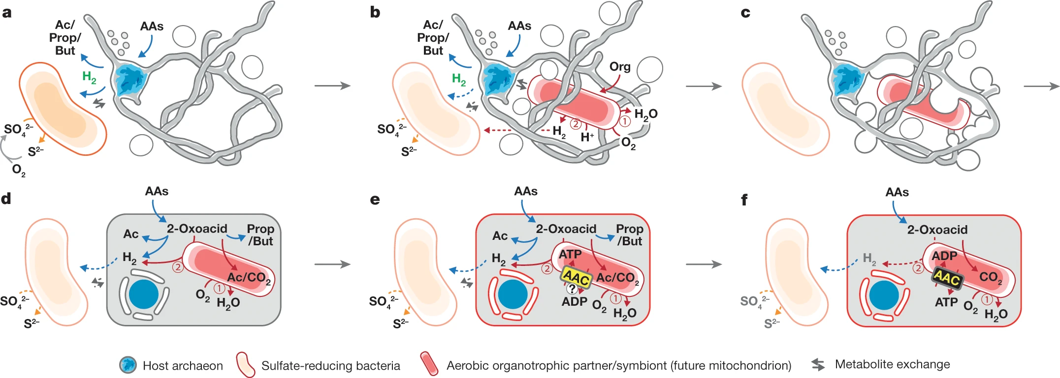
Bacteria
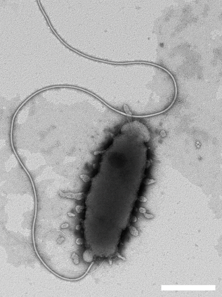
- 21 new actinobacteria: Streptomyces paludis; Streptomyces otsuchiensis; Streptomyces roseicoloratus; Gryllotalpicola protaetiae; Nonomuraea deserti, Nonomuraea diastatica, Nonomuraea longispora, Nonomuraea mesophila; Paraconexibacter algicola; Nakamurella flava; Nesterenkonia salmonea, Nesterenkonia sphaerica; Cryptosporangium phraense; Microbispora catharanthi; Serinibacter arcticus; Naasia lichenicola; Tsukamurella asaccharolytica, Tsukamurella conjunctivitidis, Tsukamurella sputi; Desertihabitans brevis; Corynebacterium sanguinis;
- 13 new bacterioids: Faecalibacter macacae; Maribacter algicola; Pleomorphovibrio marinus; Flavobacterium psychrotolerans; Flavobacterium microcysteis; Gramella sabulilitoris; Seonamhaeicola maritimus; Flavicella sediminum; Formosa maritima; Arenibacter aquaticus; Dyadobacter flavalbus; Tamlana fucoidanivorans; Thermaurantimonas aggregans;
- 2 new chloroflexes: Ktedonosporobacter rubrisoli; Tepidiforma bonchosmolovskayae;
- 2 new cyanobacteria: Neowollea manoromense; Nodosilinea signiensis;
- 7 new firmicutes: Erysipelothrix piscisicarius; Paenibacillus protaetiae; Paenibacillus antri, Paenibacillus mesophilus; Absicoccus porci; Sediminibacillus terrae; Gudongella oleilytica;
- 30 new proteobacteria: Asticcacaulis tiandongensis; Sphingobium fluviale; Glycocaulis profundi; Mixta tenebrionis; Altererythrobacter rhizovicinus; Paracoccus liaowanqingii; Croceicoccus sediminis; Photobacterium salinisoli; Eikenella exigua; Vogesella urethralis; Luteimonas lumbrici; Pseudidiomarina gelatinasegens; Vibrio sinensis, Vibrio viridaestus; Enterobacter wuhouensis, Enterobacter quasihormaechei; Roseovarius spongiae; Campylobacter armoricus; Pseudomonas saxonica; Rubrivivax albus; Haliea alexandrii; Nitrincola tapanii; Pseudomonas rhizoryzae; Pseudorivibacter rhizosphaerae; Shewanella maritima; Pseudooceanicola onchidii; Marinobacter fonticola; Sphingomonas populi; Mesorhizobium intechi; Halomonas borealis, Halomonas niordiana;
SARs
- 2 new oomycetes: Halophytophthora souzae, H. insularis;
- 9 new ochrophytes: Neidiomorpha dianshaniana; Gomphosinica selincuoensis; Cymbella hechiensis; Mallomonas lamii; Phaeoschizochlamys santosii, Phaeoschizochlamys siveri, Phaeothamnion wetherbeei, Stichogloea dopii, Stichogloea fawleyi;
- 2 new apicomplexans: Haemoproteus obainae, Haemoproteus deharoi;

Plants
- 2 new rhodophytes: Sporolithon amadoi; Hildenbrandia japananense;
- 1 new chlorophyte: Eudorina compacta;
- 1 new pteridophyte: Stegnogramma australis;
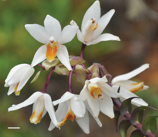
- 11 new monocots: Cymbidium atrolabium; Phragmipedium sp.; Cyrtopodium vestitum; Festuca devesae; Chascolytrum neobulbosum; Chlorospatha minima, Chlorospatha silverstonei; Piresia tenella; Habenaria gracilisegmenta; Dichorisandra striatula; Araeococcus lageniformis;
- 34 new eudicots: Euphorbia minuscula; Begonia medeiroii; Begonia handibadaganathensis; Begonia bachulkarii; Begonia itaipeensis; Tetrathylacium longirachis, T. sarayakuorum; Thesium aspermontanum, T. dmmagiae, T. neoprostratum, T. nigroperianthum, T. rhizomatum, T. stirtonii; Pinguicula simulans; Ombrophytum villamariensis; Neobartsia matuy; Aeschynomene eridesii; Lantana caudata; Atriplex turcica; Ainsliaea simplicissima; Siphocranion flavidum; Silene orientoalborzensis, S. circumcarmanica; Chrysosplenium macrospermum; Camellia debaoensis; Aeschynomene chicocesariana; Bernardia allemii; Dalechampia margarethiae; Turnera acangatinga, T. ibateguara; Fritzschia atropurpurea; Monteiroa rubra; Besleria discreta; Solanum adamantium;

Fungi
- 2 new glomeromycetes: Diversispora peloponnesiaca; Nanoglomus plukenetiae;
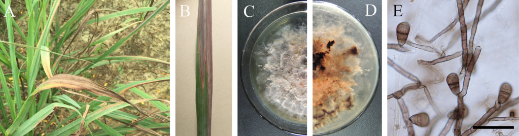
- 15 new ascomycetes: Pseudoplagiostoma myracrodruonis; Keissleriella bambusicola; Tubeufia sahyadriensis; Strigula oleistrata; Simplicillium formicae; Leptographium breviuscapum, L. conplurium, L. pseudoalbum, L. rhizoidum, L. sylvestris, L. xiningense; Saturnispora mangrovi; Saccharomycopsis oxydans; Torulaspora nypae; Aspergillus yunnanensis;
- 11 new basidiomycetes: Fomitopsis ginkgonis; Fulvifomes yoroui; Ganoderma weixiensis; Phragmidium altaicum; Trechispora yunnanensis; Amylosporus annosus; Heterocephalacria sinensis, Phaeotremella lacus, Solicoccozyma aquatica; Mycosarcoma aegyptiacum; Ochropsora staphyleae;

Sponges
- 4 new demosponges: Lissodendoryx (L.) barkleyensis, L. (L.) littoralis, L. (L.) toxaraphida, Myxilla (Myxilla) austini;
Cnidarians
- 1 new anthozoan: Trissopathes grasshoffi;
Flatworms
- 2 new polyclads: Bisacculosuteri marcelae, Paraplanocera oligoglenoides;
- 2 new rhabdocoelans: Alcha sinensis, Trigonostomum sinensis;
- 1 new triclad: Winsoria bipatria;
- 3 new monogeneans: Rhinoxenus cachorra; Biotodomella mirospinata; Calicotyle hydrolagi;
- 1 new trematode: Atriophallophorus winterbourni;
Mollusks
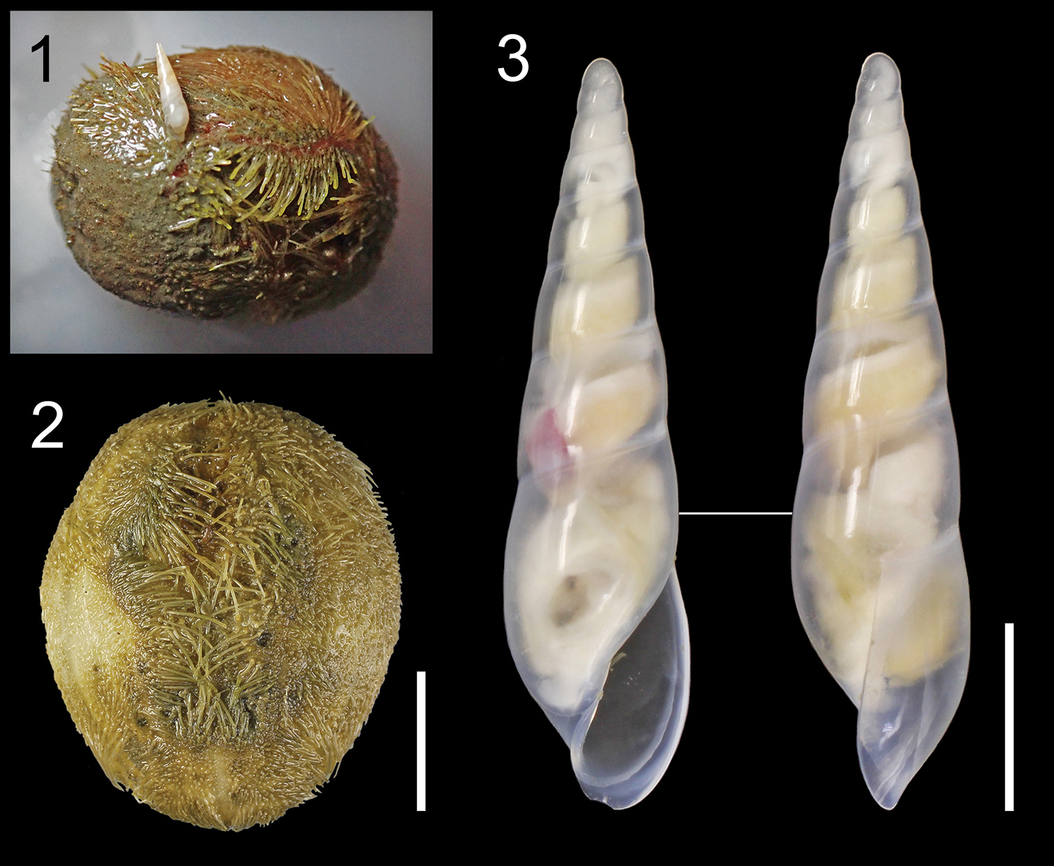
- 1 new bivalvian: Contradens novoselovi;
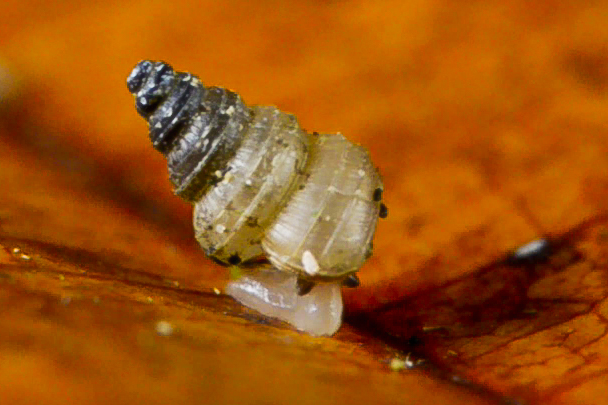
- 3 new gastropod: Onchidium melakense; Chromodoris celinae, Chromodoris helium;

Annelids
- 1 new clitellate: Graliophilus zeilensis;
Nemerteans
- 1 new species: Dushia wijnhoffae;
Nematomorphs
- 1 new species: Gordius terrestris;
Nematodes
- 5 new species: Cribronema sturhani; Perspiria boucheri; Spinitectus (Paraspinitectus) palmyraensis; Mesacanthion jejuensis; Falcaustra samarensis;
Tardigrades
- 2 new species: Milnesium cassandrae; Neoechiniscoides aski;
Arachnids
- 7 new mites: Torrenticola trigona, Monatractides hamatapodemus; Najadicola loeiensis; Phytoptipalpus occultuae; Neotetranychus longisetus; Fissicepheus parastriganovae; Paraplothrombium maketawa;
- 2 new harvestmen: Progonyleptoidellus bocaina, P. picinguaba;
- 2 new solifuges: Gylippus (Paragylippus) hakkaricus; Gaucha sp.;
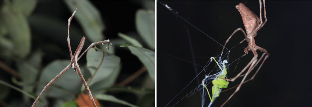
- 39 new spiders: Latrodectus umbukwane; Ischnothyreus tergeminus, Orchestina colubrina, O. zhiwui; Araneus conexus, A. digitatus; Sinoderces luohanensis, S. xueae, S. taichi, S. wenshanensis, S. aiensis, S. khanensis, S. phathaoensis, S. kieoensis, S. saraburiensis, S. dewaroopensis; Coddingtonia erhuan, C. lizu, C. huifengi; Heptathela kojim, H. sumiyo, H. uken, H. aha, H. gayozan, H. kubayama, H. mae, H. otoha, H. shuri, H. tokashiki, H. unten, H. crypta; Amilaps mayana; Loxosceles tenochtitlan; Tmesiphantes amazonicus, T. guayarus, T. nordestinus, T. raulseixasi; Kut izmiricus, K. dimensis;

Myiapods
- 11 new chilopods: Schendylops janelao; Aphilodon caboclos, A. indespectus, A. meganae, A. pereirai, A. silvestrii, Mecophilus tupiniquim, Mairata butantan, M. itatiaiensis; Lithobius (Ezembius) hualongensis, Lithobius (Ezembius) sui;
- 6 new diplopods: Polydesmus biscayensis, P. asturiensis; Bactrodesmus grandis, Hemisphaeroparia longibrachiata, H. avis, Physetoparia complexa;
Crustaceans
- 1 new cirripede: Pyrgoma spurtruncata;
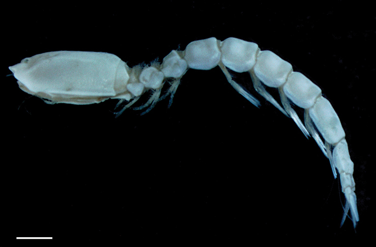
- 11 new amphipods: Crangonyx ephemerus, Crangonyx pseudoephemerus; Hyalella cheyennis; Galeatylus coripes; Exiliphotis petila, Latigammaropsis careocavata, Photis bronca, Photis posterolobus, Photis longicarpus, Podoceropsis insinuomanus, Podoceropsis pseudoclavapes;
- 2 new isopods: Bambalocra intwala; Gnathia bermudensis;
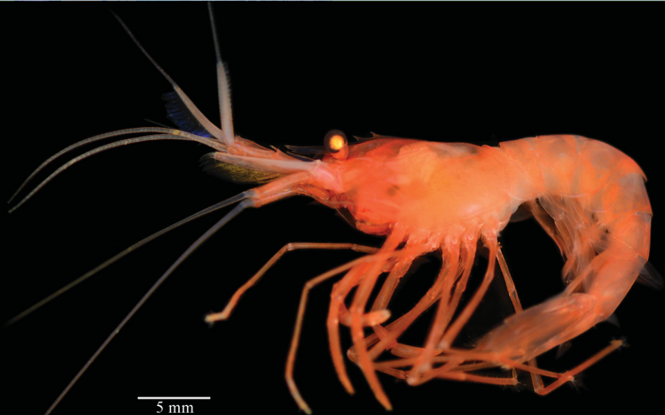
- 5 new decapods: Iridopagurus martinvaz, Nematopagurus micheleae, Pagurus carmineus; Caridina biyiga; Munidopsis spinifrons;
Hexapods
- 3 new collembolans: Arrhopalites mendoncae, Pararrhopalites queirozi, Coecobrya phoenix;
- 1 new odonate: Amphicnemis rigiketit;
- 3 new ephemeropterans: Miroculis wolverine, M. samba; Diamantina ulmeri;
- 2 new dictyopterans: Acorhinotermes claritae; Rhynchotermes armatus;
- 5 new orthopterans: Vescelia dulcis; Neocyrtopsis (Neocyrtopsis) shimianensis, Doicholobosa acuta; Anaulacomera trispinata; Cacoplistes (Laminogryllus) latioribus;
- 1 new plecopteran: Kamimuria circumspina;
- 17 new psocopterans: Aegypoecus bengalensis; Sarahcultrix ypsilophora, Sarahcultrix sphenura; Guimaraesiella corydoni, Guimaraesiella latirostris, Guimaraesiella cyanophoba, Guimaraesiella altunai, Guimaraesiella forcipata; Brueelia thorini, Brueelia straseviciusi, Brueelia mattsonae, Brueelia novemstriata, Brueelia benkmani, Brueelia arizonae, Brueelia hellstromi, Brueelia dolorosa, Brueelia melancholica;
- 5 new thysanopterans: Dendrothrips kinape; Tenothrips keruing; Anaphothrips nonporous, A. oroqeni, A. qinghaiensis;
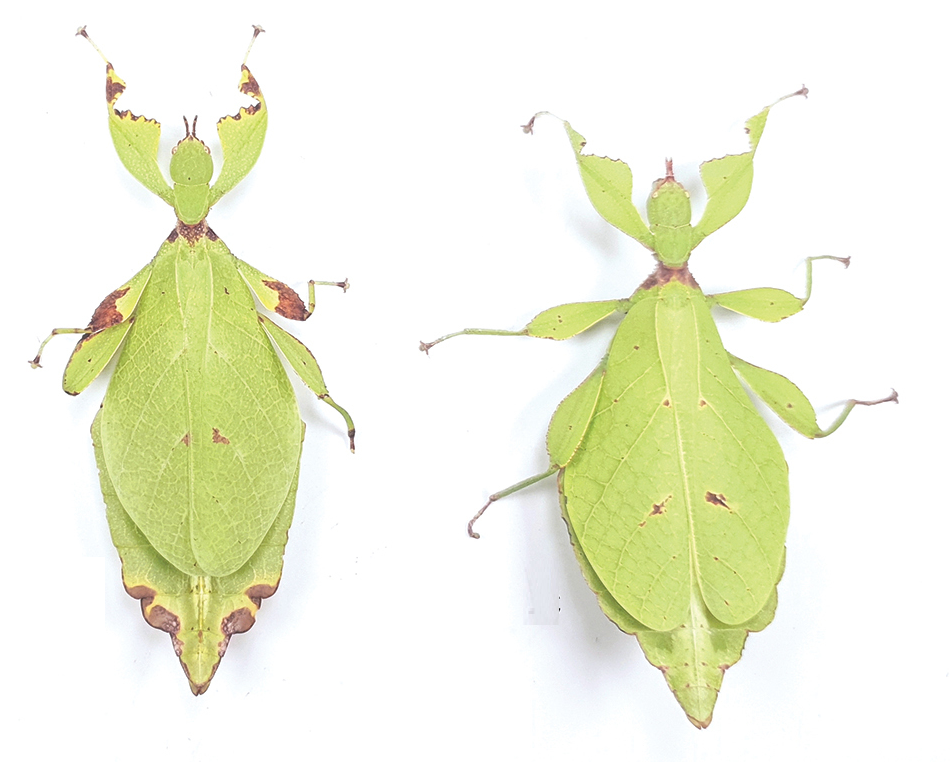
- 10 new hemipterans: Sphodrembas fumipennis, Ectomocoris caccabatus; Myxia belinda; Sinocentrus brevicornis; Longicornus brevispinus; Devolana tuxcacuensis, D. youajla, D. xajxayakamej; Bajacanthus immaculatus; Thysanaspis dennorum;
- 2 new neuropterans: Ungla pseudomeleoma; Ceraeochrysa tacanensis;
- 2 new strepsipterans: Halictophagus crassiartus, Coriophagus hansoni;
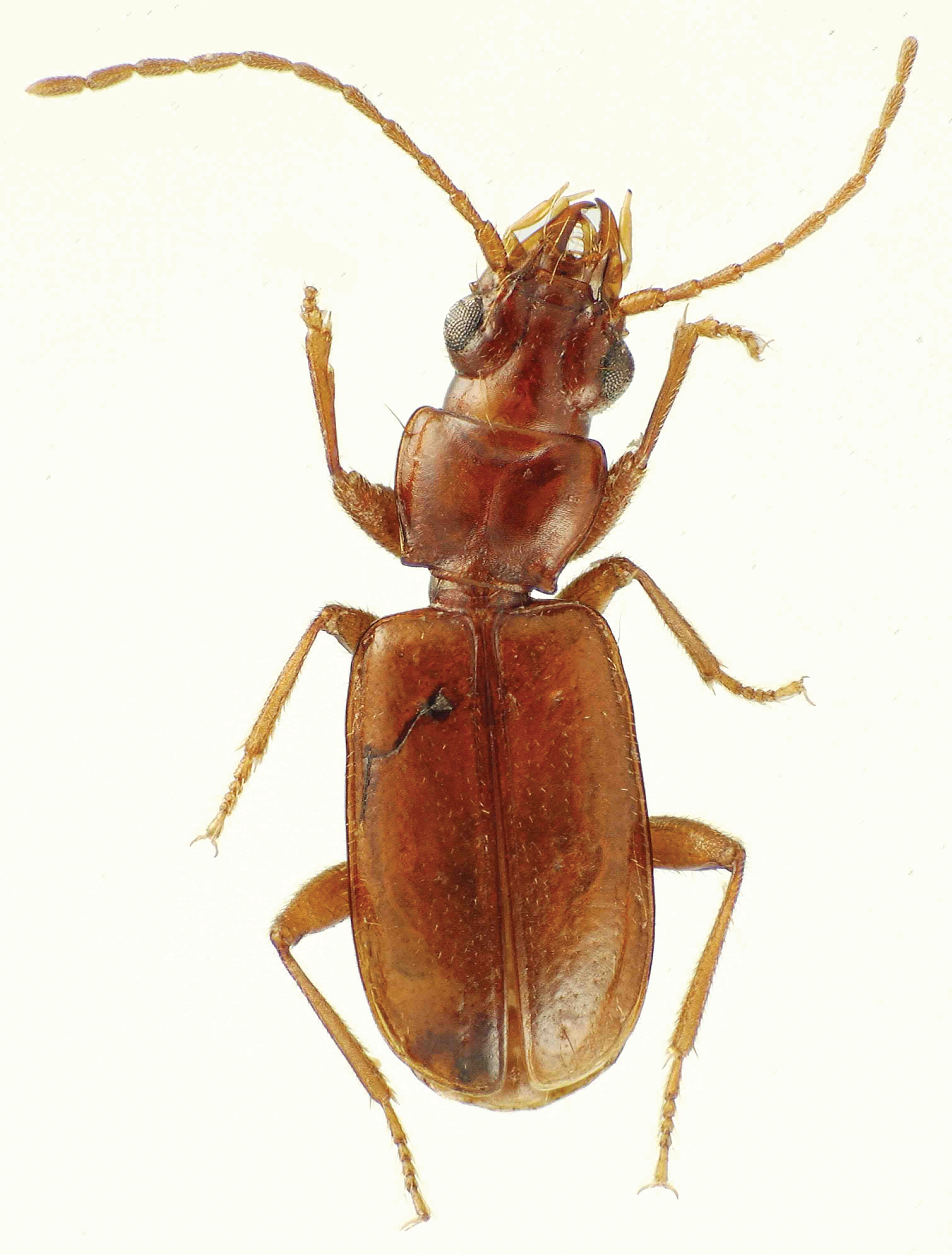
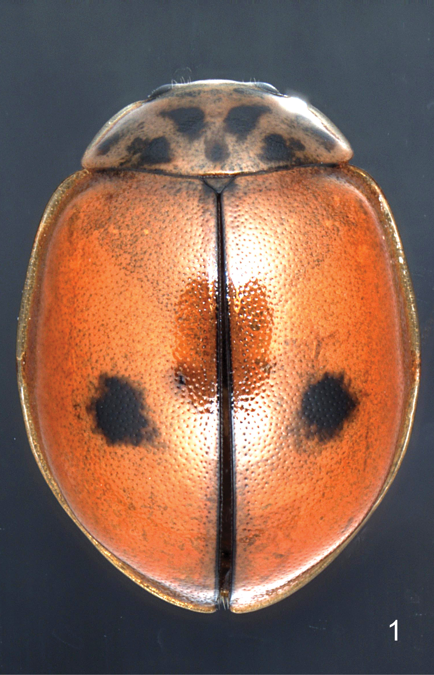
- 101 new coleopterans: Vandenbergheius celaquensis, Adetus croton, Estoloides sinaloana; Dorcus tianlongi; Deronectes kabilcevz, Deronectes propedoriae; Gnaptorina (G.) acutangula, G. (G.) crassitibia; Hygrogeus chinensis; Neoscelis hexakrotes; Colacus rubrofemoratus; Pseudopyrochroa grzymalae; Lepanus burnetti, Lepanus oxleyi, Lepanus eungella, Lepanus dukungarri,Lepanus geoffi, Lepanus yorkensis, Lepanus reidi, Lepanus topend, Lepanus pseudovillosus, Lepanus reticulatus; Barclayanthus micros, B. cooteri, Janakius sylvaticus; Onthophagus chimalapensis; Chaetocnema angustifrons, C. appendiculata, C. baoshanica, C. dapitanica, C. glabra, C. greenica, C. jinxiuensis, C. hongkongensis, C. latapronota, C. midimpunctata, C. nigrilata, C. parafusiformis, C. paragreenica, C. paraumesaoi, C. purerulea, C. reteimpunctata, C. sabahensis, C. subbasalis, C. trapezoida; Paratrichius adornatus, Paratrichius tergorufus, Paratrichius zonatus; Berosus illuviosus, B. parvus, B. andreazzei; Berosus illuviosus, B. parvus, B. andreazzei; Cheilonycha bucephalauripennis; Tonkinaphaenops yinquanicus, T. jingxicus, T. anthonyi, T. impunctatus; Veraphis loebli, V. mutsuensis, V. aomoriensis, V. sakaii; Anthaxia (Merocratus) angustata, A. (M.) priska, A. (M.) violaceidorsis, A. (M.) lucifera; Amblycerus goianiensis; Morimotanthribus leigongshani; Croscherichia armass; Logasa newtoni, Logasa thayerae, Logasa comforti; Cyrtomorphus rufobrunneus; Trigonopterus atuf, T. kumbang, T. laratensis, T. porg, T. selaruensis, T. tanimbarensis, T. triradiatus; Annamanum flavimaculatum; Amamiclytus wuxingensis; Laccobius (Glyptolaccobius) motuoensis; Copelatus martinbaehri; Aegognathus dulima, A. arnaudi; Liogenys acuta, L. amazonica, L. angustitarsis, L. clinocarinata, L. crassopunctata, L. hirsuta, L. latoemarginata, L. leviscutata, L. ornativentris, L. rodriguesi, L. pruinosa, L. quadrata, L. susanalbertorum, L. variabilis, L. vazdemelloi;
- 154 new hymenopterans: Monoctonus brachyradius, M. canadensis, M. inexpectatus, M. luteus, M. parvipalpus; Metanteon poirieri; Myiocephalus zwakhalsi; Rhathymus atitlanicus; Wesmaelia longius; Pambolus (Phaenodus) shujai, Parachremylus trachysi; Centistidea acrocercopsi, C. cosmopteryxi, C. tihamica; Glyptapanteles alejandrovalerioi, G. alexborisenkoi, G. alvarowillei, G. andybennetti, G. andydeansi, G. annettewalkerae, G. barneyburksi, G. billbrowni, G. bobhanneri, G. bobkulai, G. bobwhartoni, G. boharti, G. brianestjaquesae, G. carlhuffakeri, G. carlossarmientoi, G. carlrettenmeyeri, G. charlesmicheneri, G. charlesporteri, G. chrisdarlingi, G. chrisgrinteri, G. christerhanssoni, G. corriemoreauae, G. daveroubiki, G. daveschindeli, G. davesmithi, G. davidwahli, G. donquickei, G. eowilsoni, G. garygibsoni, G. gavinbroadi, G. gerarddelvarei, G. henrytownesi, G. howelldalyi, G. hugokonsi, G. iangauldi, G. ianyarrowi, G. ilarisaaksjarvi, G. jacklonginoi, G. jamesrobertsoni, G. jeremydewaardi, G. jesusugaldei, G. jjrodriguezae, G. johnburnsi, G. johnheratyi, G. johnlasallei, G. johnnoyesi, G. lubomasneri, G. malloryvanwyngaardenae, G. marjorietownesae, G. markshawi, G. meganmiltonae, G. mehrdadhajibabaei, G. mikegatesi, G. mikeschauffi, G. mikesharkeyi, G. nataliaivanovae, G. nealweberi, G. ninazitaniae, G. pamitchellae, G. paulhansoni, G. paulheberti, G. paulhurdi, G. philwardi, G. robbinthorpi, G. ronaldzunigai, G. roysnellingi, G. scottmilleri, G. scottshawi, G. shelbystedenfeldae, G. sondrawardae, G. stephaniecluttsae, G. stephaniekirkae, G. sujeevanratnasinghami, G. sureshnaiki, G. sydneycameronae, G. tanyadapkeyae, G. victoriapookae, G. wonyoungchoi, G. alexwildi, G. andrewdebeveci, G. andysuarezi, G. andywarreni, G. ankitaguptae, G. betogarciai, G. carinachicaizae, G. celsoazevedoi, G. claudiamartinezae, G. diegocamposi, G. dorislagosae, G. edgardpalacioi, G. edwinnarvaezi, G. erictepei, G. felipesotoi, G. ferfernandezi, G. genorodriguezae, G. grantgentryi, G. gunnarbrehmi, G. haroldgreeneyi, G. helmuthaguirrei, G. henryhespenheidei, G. jaquioconnorae, G. jerrypowelli, G. jimmilleri, G. johnstiremani, G. josesimbanai, G. juanvargasi, G. jumamuturii, G. keithwillmotti, G. kevinjohnsoni, G. kyleparksi, G. linghsiuae, G. luchosalagajei, G. malleyneae, G. mamiae, G. marcelotavaresi, G. marcepsteini, G. marcpolleti, G. marshawheelerae, G. mayberenbaumae, G. michelleduennesae, G. mikepoguei, G. montywoodi, G. pachopinasi, G. petermarzi, G. phildevriesi, G. rafamanitioi, G. suniae, G. suzannegreenae, G. taniaariasae, G. thibautdelsinnei, G. thomaspapei, G. toluagunbiadeae, G. tomwallai, G. wilmersimbanai, G. yalizhangae, G. yanayacuensis; Allorhoga uberlandiensis; Xenochlora meridionalis; Zadiprion jeffreyi; Heteroperreyia kava;
- 1 new mecopteran: Bittacus vazdemelloi;
- 16 new dipterans: Maietta dextra, M. edwardsi, M. sinistra; Wilcoxia apache, W. flavipennis, W. forbesi; Dasyhelea aliciae, D. recurva, D. latiala, D. rhopaloparamera, D. cyrtostyla, D. curacaoensis; Anabarhynchus sigatoka; Pealius satakshiae; Megalothoraca rosalyae; Carcelia ethillamima;
- 12 new trichopterans: Costatrichia inaequalis, Oxyethira carajas; Neotrichia caboca, Neotrichia capitiana, Neotrichia cruviana, Neotrichia damurida, Neotrichia macuxi, Neotrichia makunaima, Neotrichia matula; Smicridea (Rhyacophylax) scalprum, Smicridea (Rhyacophylax) sororis, Smicridea (Smicridea) capao;
- 12 new lepidopterans: Plebejus chrisreai; Melanosphecia paolo; Valeria wassalberti; Daplasa nivisala; Neolycaena enkhnasani; Pedesta wangi; Lethe langsongyuni; Barsine insolita; Thitarodes shambalaensis; Gargela polyacantha, G. grandispinata; Callibryastis latiprocessus;

Chondrichthyans
- 1 new carcharhiniform: Akheilos suwartanai;
Actinopterygians
- 2 new atheriniforms: Melanotaenia wilsoni, M. sahulensis;
- 7 new characiforms: Rhinopetitia paucirastra, R. oligolepis, R. melanohumeralis, R. nigrofasciata; Moenkhausia restricta; Hyphessobrycon veredus; Characidium chicoi;
- 1 new cichliform: Thysochromis emili;
- 2 new cypriniforms: Garra magnacavus; Oreonectes guilinensis;
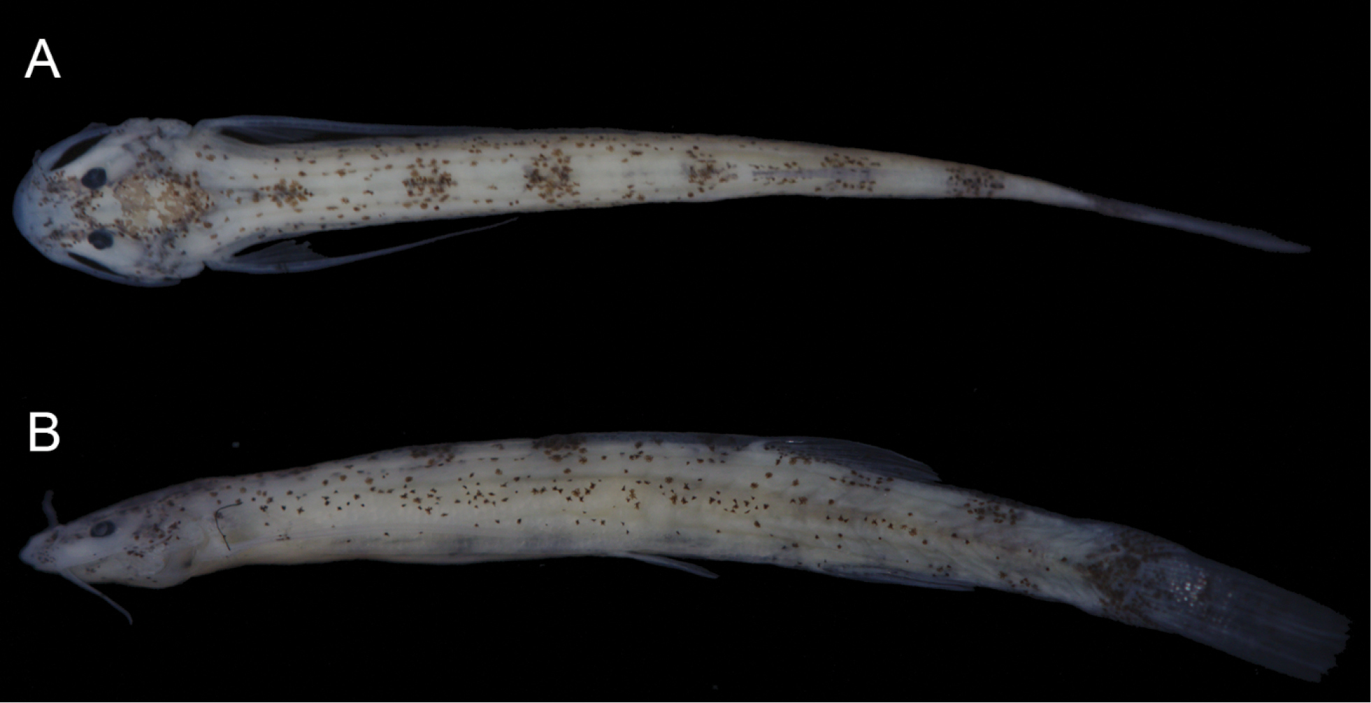
- 4 new siluriforms: Parotocinclus adamanteus; Pareuchiloglanus arcuatum, P. longicauda, P. posteranalis;
- 1 new zoarcoid: Melanostigma thalassium;
Amphibians
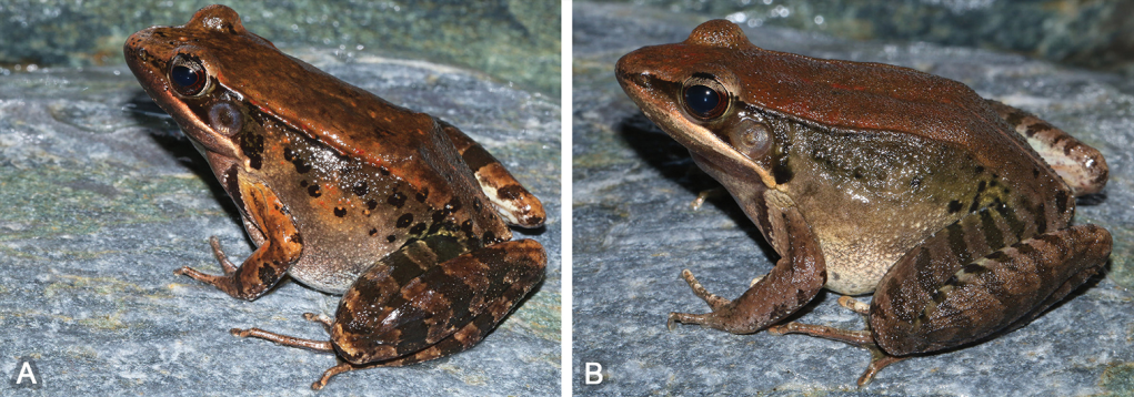
- 2 new anurans: Polypedates bengalensis; Megophrys shunhuangensis;
Reptiles
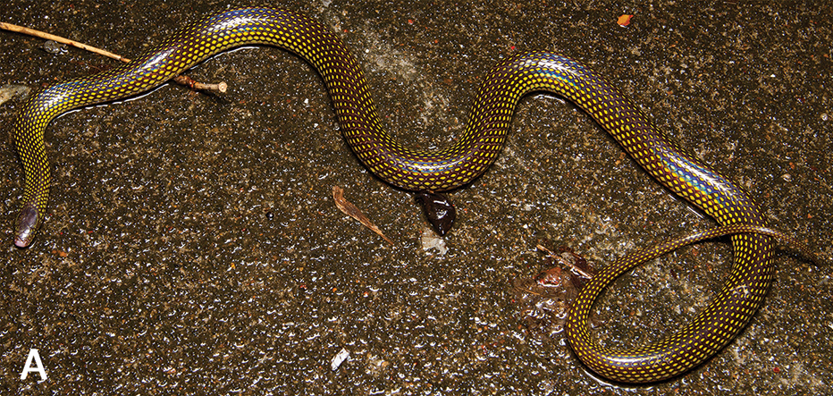
- 4 new squamates: Cyrtodactylus sp.; Cyrtodactylus muangfuangensis; Acanthosaura tongbiguanensis; Psammophylax kellyi;

– – –
* This work is licensed under a Creative Commons Attribution 4.0 International License.