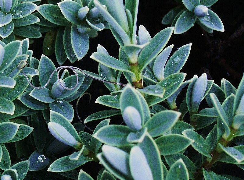
Last week I introduced the arctic willow, an unusual willow that lives as a creeping plant in the Arctic and, as I mentioned then, many species feed on this small plant. One of this species is Gynaeophora groenlandica, known as the arctic woolly bear moth.
As it is common among lepidopterans, the caterpillars of the arctic woolly bear moth feed mainly on only one species, in this case the arctic willow. But they are much more than only a caterpillar feeding on an unusual plant.
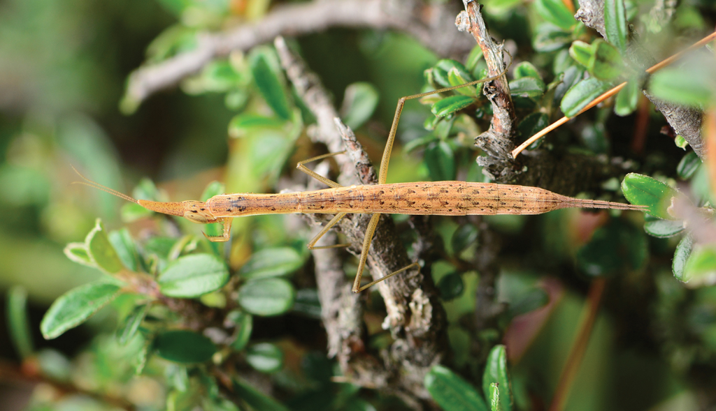
An arctic woolly bear moth among the branches of an arctic willow. Photo by Fiona Paton.*
Inhabiting greenland and the islands of Canada, the arctic woolly bear moth lives in an extreme environment in which temperatures are very low during most of the year. As a result, it is unable to remain active during several months and, like many arctic species, it hibernates.
In most of the world, the caterpillar of the arctic woolly bear moth would be considered of an average size but in its environment it is a relatevely large insect. Its body is covered by soft and long hair which varies from a reddish-brown to a dark-brown color. Adults have a grayish color with a hairy abdomen.
An adult waiting to mate. Photo by iNaturalist user pat_lorch.**
The adults mate and lay eggs around the end of June. The eggs hatch very quickly and the small first-instar larvae start to eat on arctic willow leaves but during July the temperatures start to drop quickly and the very small larvae prepare to hibernate. They spin a silken hibernaculum, a shelter to hibernate, and enter diapause, remaining inactive until June of the next year. When the snow starts to melt, they wake up, start to feed again and molt, reaching the second instar before the end of June. Then they spin another hibernaculum and enter diapause again. This cycle continues for the next years until they reach the 8th year since they hatched.
A caterpillar waking up in its hibernaculum. Photo by iNaturalist user pat_lorch.**
In that year, the caterpillars molt into pupae, which develop into adults in about a week. The adults then mate, lay their eggs, the eggs hatch and new first-instar larvae restart the cycle. Hatching in late June of the first year and mating and dying in mid June of the 8th year, the arctic woolly bear moth completes its life cycle in about 7 years, but this is restricted to 3 only weeks each year. They spend more than 90% of their life as hibernating caterpillars.
Two adults mating in June. Photo by iNaturalist user pat_lorch.**
It is not easy to be a moth in the cold Arctic. And the arctic woolly bear moth must not only survive the harsh winters but is always threatened by parasitoids, because we all know that those damn creatures exist everywhere.
And with such a specialized life cycle, what could happen with the arctic woolly bear moth now that the temperatures in the Arctic are rising? Will it survive what we have done with Earth’s climate?
Morewood WD, Ring RA (1998) Revision of the life history of the High Arctic moth Gynaephora groenlandica (Wocke) (Lepidoptera: Erebidae). Canadian Journal of Zoology 76:1371–1381.
Morewood DW, Wood MD (2002) Host utilization by Exorista thula Wood (sp. nov.) and Chetogena gelida (Coquillett) (Diptera: Tachinidae), parasitoids of arctic Gynaephora species (Lepidoptera: Lymantriidae). Polar Biology 25: 575–582. doi: 10.1007/s00300-002-0382-y
About one and a half year ago, I presented the long and thread-like wood cricket’s worm, a parasite that can control the behavior of the wood cricket and leaves its body once becoming an adult. The wood cricket’s worm belongs to the phylum Nematomorpha, commonly known as horsehair worms. They are closely related to phylum Nematoda, the roundworms. And just like horsehair worms, roundworms also love to infect crickets and grasshoppers.
One of those species is Mermis nigrescens, known as the grasshopper nematode. This worm can be found all over the world where grasshoppers exist, although they seem to be more common in Eurasia and the Americas.
An adult, gravid female. Photo by wikimedia user Beentree.**
Adults of the grasshopper nematode live in the soil and are very large for a nematode. Males measure about 5 cm in length and females can reach 20 cm, which is much larger than most nematodes that infect insects. They are, therefore, very similar to horsehair worms in appearance and behavior. The body has a smooth surface and a pale brown color, with females having a red spot on their head, the chromatopore, which functions like an eye.
After adults mate in spring or summer, males usually die but females remain in the soil through fall and winter and emerge in the following spring after a rainfall. They show a black stripe running along the body that is caused by thousands of eggs inside. They climb the nearby vegetation, up to 3 m above the ground, and lay their eggs, which measure about 0.5 mm in length, on it.
A female climbing the vegetation. Photo by Wikimedia user Notafly.**
In order to be able to climb the vegetation, female grasshopper nematodes show positive phototaxis, i.e., they are attracted by light sources, which is the opposite of what happens with most nematodes that have eyes. In fact the female’s eye, the chromatopore, is a single structure, like a single eye, and seems to have evolved independently from other nematode eyes. Its red color is caused by a hemoglobin, like the one that makes our blood red, but in this case it seems to function as a light receptor.
A closeup of the female eye and a transverse section through it. Extracted from Burr et al. (2000).*
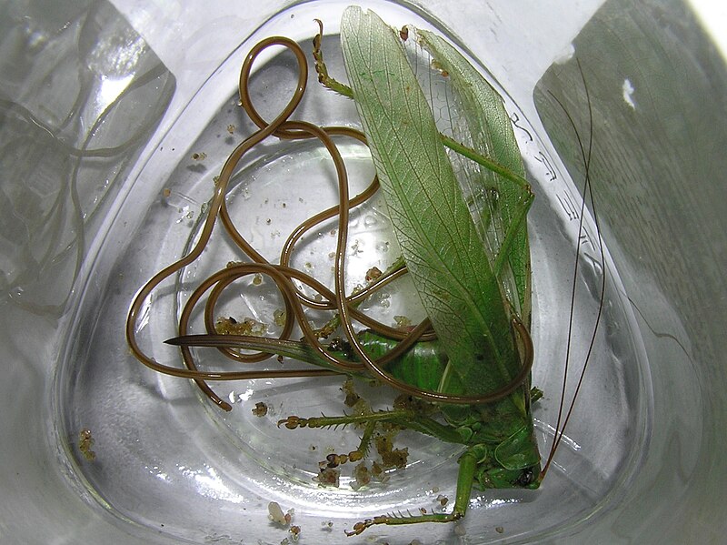
When the eggs are ingested by an orthopteran insect (usually a grasshopper but sometimes a katydid), they hatch almost immediately. The young worm pierces the grasshopper’s gut and enters its hemocoel, the “blood cavity” of the body.
An adult around its dead host, a katydid. Photo by Wikimedia user Beentree.**
There, the worm develops by absorbing nutrients from the insect’s blood directly through its cuticle. This leads to serious depletion in the insect’s levels of blood sugar, especially trehalose (the insect storage sugar) and body proteins. After reaching 5 cm or more in size, they leave the insect, killing it in the process, and continue their development in the soil until reaching the adult stage and starting the cycle all over again.
Burr AHJ, Babinzski CPF, Ward AJ (1990) Components of phototaxis of the nematode Mermis nigrescens. Journal of Comparative Physiology A 167: 245–255. doi: 10.1007/BF00188117
Burr AHJ, Hunt P, Wagar DR, Dewilde S, Blaxter ML, Vanfleteren JR, Moens L (2000) A Hemoglobin with an Optical Function. Journal of Biological Chemistry 275: 4810–4815. doi: 10.1074/jbc.275.7.4810
Burr AHJ, Schiefke R, Bollerup G (1975) Properties of a hemoglobin from the chromatrope of the nematode Mermis nigrescens. Biochimica et Biophysica Acta (BBA) – Protein Structure 405(2): 401–411. doi: 10.1016/0005-2795(75)90105-1
Gordon R, Webster JM (1971) Mermis nigrescens: Physiological relationship with its host, the adult desert locust Schistocerca gregaria. Experimental Parasitology 29(1): 66–79. doi: 10.1016/0014-4894(71)90012-9
Rutherford TA, Webster JM (1974) Transcuticular Uptake of Glucose by the Entomophilic Nematode, Mermis nigrescens. Journal of Parasitology 60(5): 804–808. doi: 10.2307/3278905
Rutherford TA, Webster JM (1978) Some effects of Mermis nigrescens on the hemolymph of Schistocerca gregaria. Canadian Journal of Zoology 56(2): 339–347. doi: 10.1139/z78-046
Japanese sea waters can emit a beautiful blue light at night, especially when disturbed. This phenomenon is caused by bioluminescent organism. One of the most famous species that produce light in the sea is the sea sparkle, a dinoflagellate. Here, however, the light is caused by a crustacean, the ostracod Vargula hilgendorfii, known in Japan as 海蛍 (umihotaru, literally “sea firefly”). Thus, the name sea firefly is commonly used to refer to the bioluminescent ostracods of the genus Vargula, most of which live along the North American Pacific coast and the Caribbbean, with Vargula hilgendorfii being the only species to occur in Japan.
Liquid light on a Japanese beach. Photo by Tdub Photo extracted from Kobi Lighting Studio.
As all ostracods, the Japanese firefly is a very small crustacean. Their body measure about 2–3 mm in length and have proportionally large eyes with about 0.2 mm in diameter. They live in the sandy substrate of shallow waters up to 5 m deep, being, therefore, benthonic, and have nocturnal habits.
A male Japanese sea firefly. The black spot is the eye. Extracted from Ogoh & Ohmiya (2005).
The areas where the Japanese sea firefly is found are marked by very strong currents. Since they have a very poor swimming ability, they are not very good to disperse to new areas but at least are able to swim well enough to avoid being carried far away by the currents. Nevertheless, the strong Japan Current seems to have slowly moved the species northward since the last ice age.
Female Japanese sea fireflies are slightly larger than males. When they copulate, they pair by touching their ventral sides while lying in opposite directions, kind of like a yin yang. Being an ovoviviparous species, the eggs remain inside the mother until they hatch, so that the female gives birth to live juveniles. Newborns are much smaller than adults, of course, but otherwise behave exactly like them, so that this species does not have a planktonic larval stage like most crustaceans.
Individual animals glowing on the sand. Photo by Tdub Photo extracted from Kobi Lighting Studio.
The diet of the Japanese sea firefly is composed mainly of debris of all sorts, including dead animals. One of the easiest ways to capture them is simply by placing dead fish as traps in the water at night and waiting for them to come.
When the Japanese sea firefly is disturbed or attacked, it releases a luminous blue cloud. This happens by the ejection of two compounds, the substrate luciferin and its enzyme, luciferase. Luciferase catalyzes the reaction of luciferin with molecular oxygen, which emits the characteristic blue light. There is a reflecting organ near the posterior end of the animal that seems to increase the power of the emitted light, which may improve their ability to escape from predators.
The natural mirror in the Japanese sea firefly’s body can enhance its light. Extracted from Abe et al. (2000).
Luciferase enzymes are of research interest especially as reporter genes. When scientists want to know whether a specific gene is being expressed in an organism, they can attach another gene right after it so that this gene will necessarily be expressed together with the gene of interest. The luciferase of the Japanese sea firefly, usually named Vargula luciferase, has been studied as a reporter gene in mammals. After creating the chain formed by the gene of interest + luciferase and inserting it into a cell, one can know whether the gene is being expressed by applying luciferin to the cells. If they glow blue this means that the gene is indeed being expressed.
As you can see, even a tiny sea creature can have a profound influence on scientific research.
Abe K, Ono T, Yamada K, Yamamura N, Ikuta K (2000) Multifunctions of the upper lip and a ventral reflecting organ in a bioluminescent ostracod Vargula hilgendorfii (Müller, 1890). Hydrobiologia 419: 73–82. doi: 10.1023/A:1003998327116
Abe K, Vannier J (1995) Functional morphology and significance of the circulatory system of Ostracoda, exemplified by Vargula hilgendorfii (Myodocopida). Marine Biology 124: 51–58. doi: 10.1007/BF00349146
Maeda Y, Ueda H, Hara T, Kazami J, Kawano G, Suzuki E, Nagamune T (1996) Expression of a Bifunctional Chimeric Protein A-Vargula hilgendorfii Luciferase in Mammalian Cells. BioTechniques 20: 116–121. doi: 10.2144/96201rr01
Ogoh K, Ohmiya Y (2005) Biogeography of Luminous Marine Ostracod Driven Irreversibly by the Japan Current. Molecular Biology and Evolution 22(7): 1543–1545. doi: 10.1093/molbev/msi155
Thompson EM, Nagata S, Tsuji FI (1989) Cloning and expression of cDNA for the luciferase from the marine ostracod Vargula hilgendorfii. PNAS 86(17): 6567–6571. doi: 10.1073/pnas.86.17.6567
Thompson EM, Nagata S, Tsuji FI (1990) Vargula hilgendorfii luciferase: a secreted reporter enzyme for monitoring gene expression in mammalian cells. Gene 96(2): 257–262. doi: 10.1016/0378-1119(90)90261-O
Vannier J, Abe K (1993) Functional Morphology and Behavior of Vargula Hilgendor Fii (Ostracoda: Myodocopida) From Japan, and Discussion of Its Crustacean Ectoparasites: Preliminary Results From Video Recordings. Journal of Crustacean Biology 13(1): 51–76. doi: 10.1163/193724093X00444
Here is a list of species described this month. It certainly does not include all described species. You can see the list of Journals used in the survey of new species here.
It’s time to come back to the fascinating flatworms and today I decide to talk about one of the most studied species in this group even though it was only formally described 15 years ago. Its name is Macrostomum lignano, or the Lignano’s macrostomum.
Measuring 1 to 2 mm in length, the Lignano’s macrostomum belongs to the order Macrostomida, one of the basalmost flatworm groups. Its body is elongate and transparent, there are two small eyes close to the anterior end, which has a small rostrum (“snout”). The mouth is a little behind the rostrum. The posterior end is broad, forming a tail plate with many adhesive organs arranged in a U-shape.
The Lignano’s macrostomum was first collected in marine samples in the city of Lignano on the Adriatic Sea coast in northern Italy in 1995 and soon revealed to be very suitable for laboratory cultures. The natural environment of this species includes the sand and other sediments near the shore. It avoids light and, when at rest, remains attached to the substrate by its tail plate. It feeds on small organism, especially diatoms, which it ingests using its cylindrical pharynx, similarly to how most flatworms eat.
Also like most flatworms, the Lignano’s macrostomum and other macrostomids have special stem cells called neoblasts that fill their body. All differentiated cells in the body come from neoblasts and are continuously replaced by them, since its differentiated cells cannot continue reproducing. Neoblasts also give the Lignano’s macrostomum an impressive regenerative ability like that of many other flatworms such as planarians.
Even before its formal description in 2005, the Lignano’s macrostomum had already been identified as a potentially new model organism. It can be easily cultured in laboratory in Petri dishes and fed with diatoms. Its body has about 25,000 cells, which is a number small enough to facilitate studies on development, regeneration, ageing and gene expression and that is exactly what has been done in the past decades.
The Lignano’s macrostomum is hermaphrodite. The body contains two testes and two ovaries. The male copulatory apparatus contains a stylet, a hardened penis-like copulatory organ. When two macrostomums mate, they touch their ventral surfaces in a yin yang fashion (just like the guys from last week) and exchange sperm. This behavior is easily observed in laboratory and led the Lignano’s macrostomum to become a model organism for the study of sexual selection as well. But wait! Sexual selection in a hermaphrodite organism? Yes! I discussed this topic some time ago here.
Two Lignano’s macrostomums mating in the yin yang position. Photo by Lukas Schärer.***
Sometimes, when two macrostomums meet, they don’t find their partner that attractive, so having their eggs fertilized by that guy is not of their interest from the female side. However, their male side is still as male as any other and they want to fertilize as many eggs as possible. As a result, if the partner is not good enough, they still want it as a female but not as a male. The other guys is not interesting in being a female only though, so copulation only occurs if both partners accept to receive each other sperm. “I let you fertilize my eggs if you let me fertilize yours.” So that’s what they do.
A pair of flatworms, Macrostomum lignano, mating. See how the white one, in the end, bends over itself and sucks the other guy’s sperm out of the female pore in order to get rid of it. Notice, however, in the last drawing, that the sperm cells are still attached to the female pore. It did not work. Image extracted from Schärer et al. (2004).
However, after they delivered the sperm into each other’s body, they separate and may never see each other again. So the female side evolved a strategy to select better sperm. When the “bad partner” moves away, a macrostomum that received low-quality sperm bends over itself, connects its pharynx to its female genital pore, and sucks the other guy’s sperm out before it has the chance to fertilize its eggs. A clever strategy, right? But remember: just as this guy is getting rid of the other guy’s sperm, the other guy may be doing the same with this guy’s sperm. So a strategy must evolve to prevent the female personality to discard the sperm. And that is exactly what happened! The sperm cells of the Lignano’s macrostomum have hard bristles pointing backward that, when the cells is pulled back, enter the tissues in the female copulatory apparatus and remain stuck. Trying to pull them out is just like trying to pull porcupine quills out of the skin.
Watch the behavior in video.
Now the male side recovered the advantage that the female side would have if the bristles were not there. But this is evolution, and its effect on hermaphrodites is like having two different personalities fighting each other in the same body.
Egger B, Ladurner P, Nimeth K, Gschwentner R, Rieger R (2006) The regeneration capacity of the flatworm Macrostomum lignano—on repeated regeneration, rejuvenation, and the minimal size needed for regeneration. Development Genes and Evolution 216:565–577. doi: 10.1007/s00427-006-0069-4
Ladurner P, Schärer L, Salvenmoser W, Rieger RM (2005) A new model organism among the lower Bilateria and the use of digital microscopy in taxonomy of meiobenthic Platyhelminthes: Macrostomum lignano, n. sp. (Rhabditophora, Macrostomorpha). Journal of Zoological Systematics and Evolutionary Research 43(2):114–126. doi: 10.1111/j.1439-0469.2005.00299.x
Lengerer B, Pjeta R, Wunderer J et al. (2014) Biological adhesion of the flatworm Macrostomum lignano relies on a duo-gland system and is mediated by a cell type-specific intermediate filament protein. Frontiers in Zoology 11:12. doi: 10.1186/1742-9994-11-12
Mouton S, Willems M, Braeckman BP, Egger B, Ladurner P, Schärer L, Borgonie G (2009) The free-living flatworm Macrostomum lignano: A new model organism for ageing research. Experimental Gerontology 44(4):243–249. doi: 10.1016/j.exger.2008.11.007
Pfister D, De Mulder K, Hartenstein V et al. (2008) Flatworm stem cells and the germ line: Developmental and evolutionary implications of macvasa expression in Macrostomum lignano. Developmental Biology 319(1):146–159. doi: 10.1016/j.ydbio.2008.02.045
Pfister D, De Mulder K, Philipp I et al. (2007) The exceptional stem cell system of Macrostomum lignano: Screening for gene expression and studying cell proliferation by hydroxyurea treatment and irradiation. Frontiers in Zoology 4:9. doi: 10.1186/1742-9994-4-9
Schärer L, Joss G, Sandner P (2004). Mating behaviour of the marine turbellarian Macrostomum sp.: these worms suck, Marine Biology145 (2) doi: 10.1007/s00227-004-1314-x
Wasik K, Gurtowski J, Zhou X et al. (2015) Genome and transcriptome of the regeneration-competent flatworm, Macrostomum lignano. PNAS 112(40):12462–12467. doi: 10.1073/pnas.1516718112
So we are going through a kind of apocalypse as everyone knows. An aggressive and contagious virus has spread all over the world and is causing a major impact in our society, killing thousands of people and crashing the economy.
But I’m not here to talk about how to protect against the virus and who is more vulnerable to it. You can find such information virtually everywhere (but don’t trust the bullshit that Karen the anti-vaxxer or your uncle Donald the boomer is spreading through Whatsapp. That is worse than the virus). Likewise, I will not point out how this pandemic is a direct outcome of our flawed capitalist society and how the fucking rich should be beheaded once and for all. No. I will make a more biological approach and explain a little bit of what this virus is from a structural, functional and taxonomic point of view.
So let’s start with what is a virus.
A virus is basically a parasitic piece of sh… genetic material that infects cells in order to reproduce. Viruses are not quite living beings as they neither have cells nor metabolism. However, they need cells to reproduce. All viruses consists of a strand of nucleic acid (either DNA or RNA) and a capsid, a “box” that protects the nucleic acid. The capsid is usually formed by many copies of one or two proteins that are encoded in the virus’ genetic material. Each individual protein molecule of the capsid is called a capsomere.
Scheme of a helical virus showing the helical capsid in green and the genetic material in blue. Credits to Anderson Brito.*
The Tobacco mosaic virus, that infects tobacco plants and others, has a typical helical capsid.
Most viruses have either a helical or an icosahedral capsid. In a helical capsid, the capsomeres are helically arranged and form an elongate and hollow tube inside of which the genetical material is located. In icosahedral capsids, the capsomeres are arranged to form a icosahedron, i.e., a polyhedron with 20 faces that surrounds the genetic material.
Scheme of an icosahedral virus with an icosahedral capsid (green) surrounding the genetic material (red). Credits to Anderson Brito.*
Adenoviruses are an example of virus with icosahedral capsids. Photo by Graham Beards.**
Many viruses have an additional coat, the envelope, that surrounds the capsid. The envelope is a bi-lipid layer crossed by glycoproteins, like the cell membrane of living organisms, and is formed by the cell membrane or an internal membrane of the cell in which the virus was born. It is, therefore, very similar to the cell membrane of the virus’ host.
Scheme of an enveloped icosahedral virus. The bi-lipid layer is shown in gray and the glycoproteins in orange. Credits to Anderson Brito.*
Zika virus (digitally colored blue in this electron microphotograph) is an envoloped icosahedral virus.
Regarding the type of nucleic acid found in viruses, they can be classified into three main groups: DNA viruses, RNA viruses and retroviruses.
DNA viruses have DNA as their nucleic acid. When they infect a cell, they are delivered into the cell’s nucleus, where they depend entirely on the cell’s machinery to reproduce, i.e., they use the hosts DNA-polymerase to produce new DNA strands and the host’s RNA-polymerase to build a viral RNA that will, in turn, be converted into the capsid proteins using the cell’s ribosomes. DNA viruses suffer little mutation because DNA-polymerase enzymes have a proofreading ability, i.e., they can detect errors during replication and fix them. Viruses such as Herpesvirus (which cause herpes and chicken pox), Poxvirus (which include the now extinct Variola virus that caused smallpox) and Adenovirus are all DNA viruses.
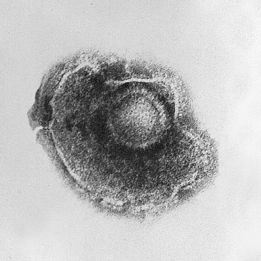
The Human alphaherpesvirus 3 (HHV-3) is an enveloped icosahedral DNA virus that causes chickenpox and shingles in humans.
RNA viruses, also called riboviruses, on the other hand, have RNA as their nucleic acid. When they infect a cell, they usually remain in the cell’s cytoplasm. Different from DNA viruses, RNA viruses often have their own RNA-polymerase enzyme and use it to produce new copies but still depend on the host’s ribosomes to translate their RNA into proteins to build the capsid and make new copies of their RNA polymerase. Since RNA-polymerase enzymes lack the proofreading ability of DNA-polymerase, RNA viruses mutate rapidly. A lot of human diseases are caused by RNA viruses, incluing the common cold, influenza, ebola, yellow fever, dengue fever, Zika fever, hepatitis C, rabies, polio, measles, as well as COVID-19, caused by the current apocalypse-driving coronavirus.
The Yellow fever virus is an enveloped icosahedral RNA virus.
Retroviruses, the last virus type, also have RNA as their nucleic acid. However, different from RNA viruses, retroviruses do not produce new copies directly from their RNA using a RNA-polymerase. Instead of that, they have another type of enzyme, called reverse transcriptase, that builds a DNA strain from their RNA. This viral DNA is then incorporated into the DNA of the host cell by an integrase enzyme. Retroviruses, therefore, change the host’s genome, i.e., they create a “hybrid” of themselves and the host. The infected cell then transcribes the viral DNA back into RNA, making several copies that allow to virus to reproduce. The most famous retroviruses to infect humans are Human immunodeficiency virus (HIV) and Hepatitis B virus.
Human Imunodeficiency Virus 1 is an enveloped icosahedral retrovirus that causes AIDS in humans.
But now let’s focus on our current celebrity, SARS-CoV-2, colloquially known as the coronavirus. This virus, which is causing the current apocalypse, is a new strain, discovered in late 2019, of the Severe acute respiratory syndrome-related coronavirus (SARSr-CoV). The previous SARS outbreak between 2002-2004 was caused by another strain of this same species, SARS-CoV, or now often referred to as SARS-CoV-1. This virus belongs to the genus Betacoronavirus and the family Coronaviridae. All members of the family Coronaviridae are often called “coronavirus” and the currently known species infect birds and mammals.
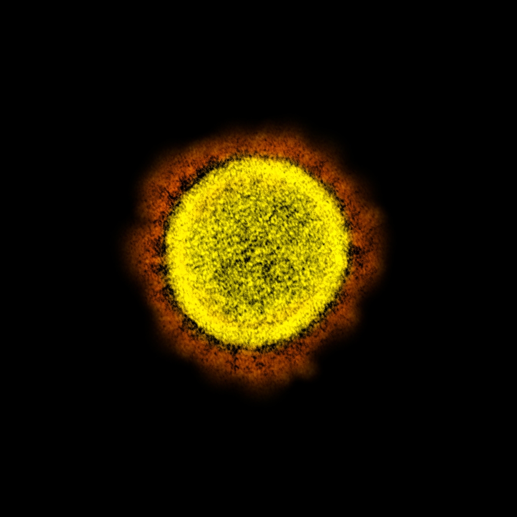
SARS-CoV-2 with artificial colors showing the “corona” (in orange) formed by the club-shaped glycoproteins of its envelope.
Coronaviruses are RNA-viruses, as mentioned above, and have a helical capsid, as well as an envelope. This envelope contains large club-shaped proteins that appear as projections on the virus surface and, in electron micrographs, resemble the solar corona, hence the name coronavirus. The envelope is built from the membrane of the host’s endoplasmic reticulum but includes glycoproteins of viral origin, including the club-shaped glycoproteins that characterize these viruses.
The presence of this envelope around the capsid has some advantages and some disadvantages to coronaviruses and any other enveloped virus. Since this envelope is basically a piece of the host’s cell, enveloped viruses can sneak into new hosts more easily because the immune system takes some time to recognize them as invaders since they are wearing a host’s “clothing”. On the other hand, this envelope is very fragile when exposed to the outer environment and degrades very quickly, so that the virus needs close contact of an infect host with a new host in order to spread. This is also why washing your hands with soap kills the virus so easily. If the virus were not enveloped, i.e., had only its capsid, it would be much more resistant.
The club-like glycoproteins of the viral envelope are also the responsible for the virus ability to infect. These glycoproteins connect to the angiotensin-converting enzyme 2 (ACE2), an enzyme that is found on the surface of many human cells. ACE2 is especially abundant in the lungs, which is the reason why this is the organ that suffers the most during SARS-CoV infections.
The main genera inside the family Coronaviridae are Alphacoronavirus, Betacoronavirus, Gammacoronavirus and Deltacoronavirus. Most known species of Alpha- and Betacoronavirus infect bats, so it is likely that the ancestor of these genera was originally a bat-specific virus that later mutated and acquired the ability to infect other species. All coronaviruses that infect humans belong to this two genera and include, besides SARS-CoV, MERS-CoV (which causes the Midle East Respiratory Syndrome) and several viruses that cause mild cold-like symptoms, such as HCoV-HKU1, HCoV-NL63 and HCoV-229E. Species of Gammacoronavirus infect mainly birds, although at least one species, Coronavirus HKU15, causes diarrhea in pigs. The genus Deltacoronavirus includes the Avian coronavirus (IBV), which causes infectious bronchitis in birds, and the Beluga whale coronavirus SW1, the only coronavirus known to infect a marine mammal.
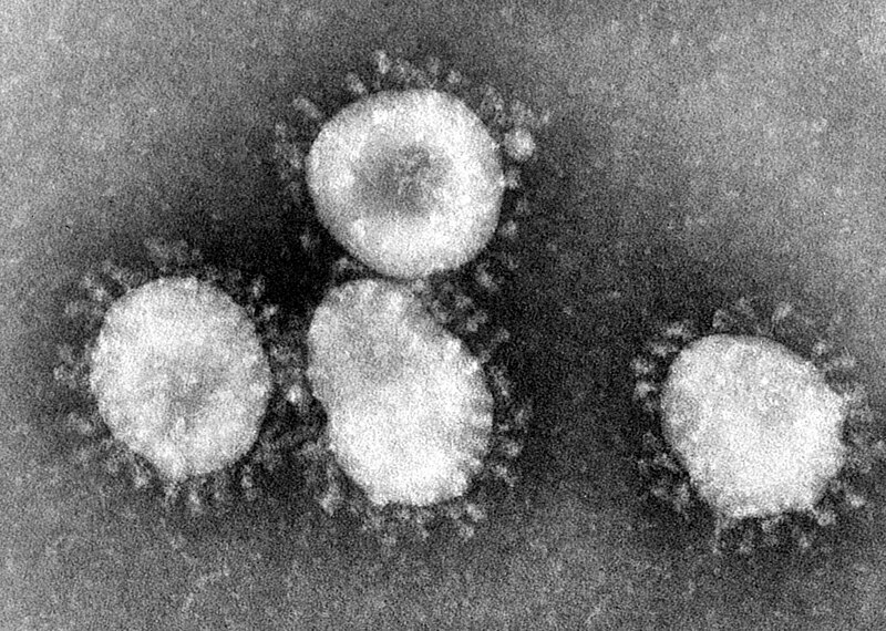
Avian coronavirus. The club-shaped glycoproteins are clearly visible on the envelopes.
The genome of coronaviruses has about 30 thousand nucleotides, being some of the largest genomes among RNA viruses. The only known RNA virus with a larger genome, with about 41 thousand nucleotides, was discovered in 2018 and infects, guess what, planarians! Named Planarian secretory cell nidovirus (PSCNV), it belongs to the order Nidovirales, which includes coronaviruses and many other RNA-viruses, but seems to have diverged from most members of Nidovirales a long long time ago. Maybe I’ll talk more about this particular virus and the implications of its discovery in a future post.
Let’s conclude this post with a quick review of what we have learned about SARS-CoV-2, the “coronavirus”:
It is an RNA-virus, meaning that it has a great mutation potential and is able to create copies of itself in the host’s cytoplasm, being an almost self-suficient virus;
It has a helical capsid surrounding its RNA;
It has an envelope derived from the membrane of the host’s endoplasmic reticulum, which is the reason why it can be so easily killed by water and soap;
This envelope includes large clube-like glycoproteins that make it appear as a solar corona in electron micrographs, hence the name coronavirus;
It is a member of the genus Betacoronavirus, which includes a lot of species known to infect bats and that’s the reason why its origin in a Chinese bat soup is very likely.
I hope that this post helped you see more about this new virus than its ability to collapse human societies.
Orchids are very popular ornamental flowers and come in a variety of colors, sizes and shapes. Finding them in natural habitats is not always that easy, but is not difficult either if you are visiting an area of Atlantic Forest in Brazil.
One orchid species that is native to this endangered biome is Cattleya coccinea, until very recently known as Sophronitis coccinea, a reason why it is still often called Sophronitis among orchid collectors. Even among collectors this species often lacks a common name, but I think “red sophronitis” fits well, although, well… almost all species in the former genus Sophronitis are red.
A specimen of Cattleya coccinea growing around the canyons near the border of the states of Santa Catarina and Rio Grande do Sul, southern Brazil. Photo by João Gava Just.*
Occurring in areas of mid to high elevations from around Espírito Santo southward to Rio Grande do Sul, Brazil, and reaching neighboring areas in Misiones, Argentina, the red sophronitis is a relatively small orchid that produces solitary flowers with a bright scarlet red color. The two petals are much broader than the three sepals, although the labellum, the third tubular petal, is narrower.
Although sometimes mentioned as an endangered species, the red sophronitis is the species in the Sophronitis group with the largest natural population and widest geographical range. One closely related species, the Mantiqueira’s sophronitis, Cattleya mantiqueirae, is in a much more critical situation and was, initially, considered a subspecies of the red sophronitis. Recent molecular analyses, though, questioned the current classification of both species and they may end up becoming again a single species or perhaps be split into more species.
The red sophronitis flowers between late winter and early spring, with the peak from August to October. The flower, normally a single one each time, is scentless and lacks nectar. Nevertheless, the conspicuous nature of the flower suggests that the plant is pollinated by a vision-oriented animal. After some observations of flowers in the wild, the only identified pollinator was the hummingbird Chlorostilbon lucidus. The small bird visits the flowers looking for nectar but, not finding any, leaves very quickly, in less than 5 seconds, but this is enough to carry pollen from one flower to another. Bees, which are common pollinators of other species of Cattleya, do not seem to have any interest in this species.
Despite its lovely appearance, the red sophronitis is a liar. It attracts a naïve pollinator promissing some reward but makes the poor creature move away with nothing. But beauty has never been a synonym of kindness despite our human efforts to think so.
Caballero-Villalobos L, Silva-Arias GA, Buzatto CR, Nervo MH, Singer RB (2017) Generalized food-deceptive pollination in four Cattleya (Orchidaceae: Laeliinae) species from Southern Brazil. Flora 234: 195–206. doi: 10.1016/j.flora.2017.07.014
Rodrigues JF, van den Berg C, Abreu AG, Novello M, Veasey EA, Oliveira GCX, Koehler S (2014) Species delimitation of Cattleya coccinea and C. mantiqueirae (Orchidaceae): insights from phylogenetic and population genetics analyses. Plant Systematics and Evolution 301:1345–1359. doi: 10.1007/s00606-014-1156-z
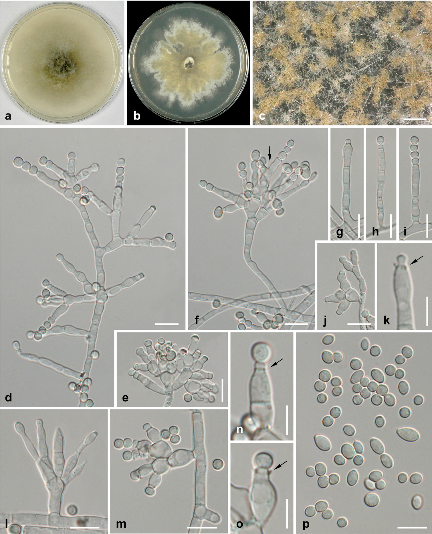
Fungi show a variety of different fruiting bodies and they are almost always named based on what those fruiting bodies look like. This is the case of today’s fellow as well. Its scientific name is Aleuria aurantia and it is commonly known as the orange peel fungus. Due to English ambiguous language, such a name could mean a fungus that grows on orange peels as well, but that is not the case, as such a species is named green mold.
The orange peel fungus is found throughout the Holarctic region, i.e., North America, Europe and Northern Asia, and eventually elsewhere, probably due to human-driven dispersal. Its main habitat includes opean areas near conifer forests, where it lives as a saprotrophe, i.e., a decomposing fungus with the mycelia scattered through the soil.
Orange peel fungus in France. Photo by David Renoult.*
The cup-shaped fruiting bodies appear more often in autumn. They have a strong orange color caused by carotenoids, especially β-carotene, γ-carotene and aleuriaxanthin. At first they are small and very regular, but grow up to 6 cm in diameter, when their structure often becomes irregular or torn apart, resembling pieces of orange peel thrown on the ground. The spores that they produce remain ungerminated in the soil until spring, when they start to grow. The cold temperatures of winter seem to be necessary for the spores to germinate, as spores in laboratory cultures only germinated successfully after remaining frozen for about 3 months.
The orange peel fungus has been a target species for pharmacological studies due to the presence of a lectin called Aleuria aurantia lectin (AAL). Lectins are a group of proteins that bind to sugars or sugar groups of other molecules such as glycoproteins and glycolipids. They are found in all groups of organisms and have different functions. In animals, for example, they are important in cell adhesion. They connect to glycoproteins on the cell membrane and tie neighbor cells to each other, preventing tissues to fall apart.
Flattened and broken fruiting bodies resembling orange peel in the United States. Credits to iNaturalist user schwee.*
In many parasitic fungi, bacteria and viruses, lectins are important to recognize the host. They also seem to have a role during the production of spores in fungi. AAL from the orange peel fungus binds to fucose, a sugar that is commonly found on the surface of cells in mammals, insects and plants as part of their glycoproteins. The name fucose comes from the fact that this sugar forms the polysaccharide fucoidan found in several species of brown algae.
AAL has several practical uses. Due to its fucose-binding behavior, it can be used to detect the presence of fucose in different cells. Fucose is also related to some types of allergies and AAL has been studied as a potential compound to develop new antiallergics.
Other than its pharmacological properties, the orange peel fungus is also edible, though not that popular as food. Its fruiting bodies can be eaten fresh or dried for storage. Due to the presence of carotenoids, the orange peel fungus has potential antioxidant properties, although studies on its nutritious value seem to be lacking.
Fukumori F, Takeuchi N, Hagiwara T, Ohbayashi H, Endo T, Kochibe N, Nagata Y, Kobata A (1990) Primary structure of a fucose-specific lectin obtained from a mushroom, Aleuria aurantia. Journal of Biochemistry 107(2):190–196. doi: 10.1093/oxfordjournals.jbchem.a123024
Ogawa S, Nagao H, Ando A, Nagata Y (2000) Enhancement of ascospore germination from Aleuria aurantia after cold storage. Mycoscience 41:287–289. doi: 10.1007/BF02489686
Ogawa S, Nakajima E, Nagao H, Ohtoshi M, Ando A, Nagata Y (1998) Synthesis of a Lectin in Both Mycelia and Fruit Bodies of the Ascomycete Mushroom Aleuria aurantia. Bioscience, Biotechnology, and Biochemistry 62(5):915–918. doi: 10.1271/bbb.62.915
Roth-Walter F, Schöll I, Untersmayr E, Fuchs R, Boltz-Nitulescu G, Weissenböck A, Scheiner O, Gabor F, Jensen-Jarolim E (2004) M cell targeting with Aleuria aurantia lectin as a novel approach for oral allergen immunotherapy. Journal of Allergy and Clinical Immunology 114(6):1361–1368. doi: 10.1016/j.jaci.2004.08.010
Seaver FJ (1914) North American Species of Aleuria and Aleurina. Mycologia 6(6):273–278. doi: 10.1080/00275514.1914.12020977
Singh U, Bhatt RP, Stephenson SL, Uniyal P, Mehmood T (2017) Wild edible mushrooms from high elevations in the Garhwal Himalaya—II. Current Research in Environmental & Applied Mycology 7(3):208–226. doi: 10.5943/cream/6/2/6
Węgiel J, Kohlmünzer S (2001) Mycelial culture of the fungus Aleuria aurantia and some of its metabolites. Pharmaceutical Biology 39(2):108–112. doi: 10.1076/phbi.39.2.108.6249
Wimmerova M, Mitchell E, Sanchez JF, Gautier C, Imberty A (2003) Crystal structure of fungal lectin six-bladed β-propeller fold and novel fucose recognition mode for Aleuria aurantia Lectin. Journal of Biological Chemistry 278: 27059-27067. doi: 10.1074/jbc.M30264220
Seven years ago I discussed the phylogenetic position of Acoelomorpha and their close relative, Xenoturbella, which together form the clade Xenacoelomorpha. Being very simple bilaterian animals, lacking almost every major structure common to most other bilaterians, their exact position is usually considered to be basal inside Bilateria but the idea that they are deuterostomes was raised after some molecular studies grouped them with the clade Ambulacraria, which includes echinoderms and hemichordates.
Being deuterostomes would mean that Xenacoelomorpha suffered a huge simplification of their anatomy. Back in 2013, when I wrote the other article, this was causing a lot of controversy but, a time after that, new molecular studies confirmed the basal position of Xenacoelomorpha and it became kind of well accepted that they are, indeed, the basalmost clade in Bilateria.
A simplified version of the animal tree of life showing the uncertain position of Xenacoelomorpha. The position of Placozoa and Ctenophora is not very clear too.
But once a trouble, always a trouble.
By 2019, a new study that tried to anticipate effects of systematic errors during molecular phylogeny studies, such as long-branch attraction, concluded that the basal position of Xenacoelomorpha is an artifact and that, when one tries to minimize the errors, their position in Deuterostomia becomes clear. As I mentioned in my old post, the main problem in Xenacoelomorpha appearing inside Deuterostomia is related to their oversimplification. They lack almost everything that any typical bilaterian has. What would have forced them to become that simple?
Another recent study suggested that, in the case of Xenoturbella at least, this may be the result of their soft-substrate burrowing habits. They compare Xenoturbella to nudibranchs, among which some species have similar lifestyles. One of these nudibranchs, Xenocratena, was actually discovered at about the same time as Xenoturbella living in the same environment. They have a paedomorphic (more simplified, “baby-like”) anatomy compared to other nudibranchs. However, it is not at all as simple as Xenoturbella.
On the other hand, there is another genus of nudibranchs that is indeed oversimplified, Pseudovermis, and it lives burrowed in soft substrate as well. Molecular analyses revealed that Pseudovermis is not closely related to Xenocratena but to Cumanotus, another burrowing nudibranch, which suggests that this simplification occurred twice among nudibranchs.
Phylogenetic relationships among nudibranchs. See Pseudovermis and Cumanotus at about 2 o’clock and Xenocratena at about 7 o’clock. Credits to Martynov et al. (2020).*
This is not an evidence that Xenoturbella is a simplified deuterostome, but it is a good argument. But what about the simplifications of Acoelomorpha? I think that if Xenoturbella was not closely related to Acoelomorpha I would be more willing to accept this hypothesis. My heart leans toward the hypothesis of basal Xenacoelomorpha, though. However, as any cientist should do, I will accept Xenacoelomorpha as deuterostomes if enough evidence is presented.
Xenoturbella is always the main problem in this equation, The nervous system of Acoelomorpha, for example, although simplified, has kind of the basic pattern found in all bilaterians and could have evolved from the oral ring in a cnidarian-like ancestor according to some hypotheses. In Xenoturbella, though, the nervous system is much weirder, being formed by a simple network of difuse neurons below their skin. I guess addressing the organization of the nervous system in all these groups is a good topic for another post.
If there is one thing, in my opinion, that makes the position of Xenacoelomorpha within Deuterostomia somewhat convincing is the fact that many features of Deuterostomia seem to be more primitive inside Bilateria when compared to those in Protostomia, so the position of Xenacoelomorpha among Deuterostomia is more plausible than their position among Protostomia(although this is not even considered possible anymore) for sure. We tend to think that deuterostomes are more “derived” simply because humans are deuterostomes. But this discussion is also a subject for another post.
What do you think? Are you team basal or team deuterostome?
Cannon JT, Vellutini BC, Smith J, Ronquist F, Jonfelius U, Hejnol A (2016) Xenacoelomorpha is the sister group to Nephrozoa. Nature 530: 89–93. doi: 10.1038/nature16520
Jondelius U, Raikova OI, Martinez P (2019) Xenacoelomorpha, a Key Group to Understand Bilaterian Evolution: Morphological and Molecular Perspectives. In: Pontarotti P (ed) Evolution, Origin of Life, Concepts and Methods. Cham: Springer International Publishing, . pp. 287–315. doi: 10.1007/978-3-030-30363-1_14
Martynov A, Lundin K, Picton B, Fletcher K, Malmberg K, Korshunova T (2020) Multiple paedomorphic lineages of soft-substrate burrowing invertebrates: parallels in the origin of Xenocratena and Xenoturbella. PLoS ONE 15(1): e0227173. doi: 10.1371/journal.pone.0227173
Philippe H, Poustka AJ, Chiodin M, et al. (2019) Mitigating Anticipated Effects of Systematic Errors Supports Sister-Group Relationship between Xenacoelomorpha and Ambulacraria. Current Biology 29(11):1818–1826. doi: 10.1016/j.cub.2019.04.009
People love to name sea creatures by making analogies with things found on land. Today’s species is one more of this type, being the best known species of the so-called sea butterflies and, therefore, known as the common sea butterfly. It has nothing to do with butterflies, though, and its scientific name, Limacina helicina, is better to describe it.
The common sea butterfly is a mollusk, more precisely a gastropod, and, as it has a shell, it is a “snail”. It does not crawl through the floor as most snails though. With a spiral shell measuring only about 10 mm in diameter, it lives in the water column and is sometimes described as a planctonic species. It can swim by itself, though, because its fleshy foot is changed into two expansions called parapodia that act as two large fins. Its shell is mostly transparent and the soft parts are mainly purple, although the parapodia are almost transparent as well.
The common sea butterfly is truly a beautiful creature, isn’t it? Photo by Russ Hopcroft, University of Alaska, Fairbanks.
The habitat of the common sea butterfly includes the cold waters of the Arctic region, including the Arctic Ocean and neighboring areas of the Atlantic and Pacific oceans. In the Pacific, it can occur southward to Japan and the northern parts of the United States. Larger specimens tend to inhabit deeper waters, up to 150 m deep, while smaller ones live closer to the surface, up to 50 m down. Until very recently, the common sea butterfly was thought to inhabit Antarctic waters as well but molecular studies revealed that the populations around Antarctica belong to a different species, Limacina antarctica.
See how fast they can beat their wings.
The diet of the common sea butterfly includes several smaller planktonic creatures, especially small crustaceans, such as nauplii (larvae) of copepods, as well as dinoflagellates, ciliates and diatoms. Juveniles of their own species are also very common, sometines making up the second most common item in their diet. In order to capture food, they produce a spherical web of mucus that floats above them in the water. This web traps other organisms in the water column and is later sucked and eaten by the sea butterfly together with the entrapped creatures. This web is very difficult to observe during the day because of diffuse refraction but appears clearly at night. When disturbed by light, however, the common sea butterfly tends to quickly swallow its web and sink to escape from danger.
A commo sea butterfly with its spherical mucous web seen as an oval concentration of finer particles right above the snail. Extracted from Gilmer & Harbinson (1986).
The thin shell of the common sea butterfly consists of aragonite, which is highly soluble and sensitive to changes in temperature and acidification of the water. Studies have shown that the expected increase in ocean acidification due to human-induced climate changes will probably have a negative impact on populations of the common sea butterfly and related species. This is particularly worrisome regarding the common sea butterfly because it is the a key species in Arctic ecosystems, being an important food source for many marine animals, such as fish, whales, birds and even other mollusks.
The little snail will not give up the fight so easily, though. Studies have shown that the periostracum (the outer organic layer of the shell) can prevent the aragonite to dissolve and an physical trauma that breaks the periostracum, allowing the direct contact of the aragonite with the water, is necessary to cause dissolution. And even when this happens, the common sea butterfly can compensate by building new aragonite layers on the inner surface of the shell and it is able to extract aragonite from water for this purpose even when the levels in water are very low.
The common sea butterfly is small but it is also tough.
Corneau S, Alliouane S, Gattuso JP (2012) Effects of ocean acidification on overwintering juvenile Arctic pteropods Limacina helicina. Marine Ecology Progress Series 456:279–284. doi: 10.3354/meps09696
Comeau S, Jeffree R, Teyssié JL, Gattuso JP (2010) Response of the Arctic pteropod Limacina helicina to projected future environmental conditions. PLoS One 5(6):e11362. doi: 10.1371/journal.pone.0011362
Gilmer RW, Harbinson GR (1986) Morphology and field behavior of pteropod molluscs: feeding methods in the families Cavoliniidae, Limacinidae and Peraclididae (Gastropoda: Thecosomata). Marine Biology 91:47–57. doi: 10.1007/BF00397570
Lischka S, Büdenbender J, Boxhammer T, Riebesell U (2011) Impact of ocean acidification and elevated temperatures on early juveniles of the polar shelled pteropod Limacina helicina: mortality, shell degradation, and shell growth. Biogeosciences 8:919–932. doi: 10.5194/bg-8-919-2011
Last week I presented a beautiful sea snail, the common sea butterfly, with wing-like parapodia that allows it to swim. The sea butterflies belong to a group of marine gastropods known as Pteropoda due to this foot modified into fins. There are two main groups of pteropods, Thecosomata, that have a shell, and Gymnosomata, that don’t have a shell. While the shelled ones are called sea butterflies, the naked ones are called sea angels or naked sea butterflies.
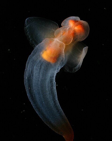
The most popular sea angel is Clione limacina, the common sea angel. Its body is mostly transparent and, like all pteropods, has two parapodia that look like wings which, together with its elongate and shell-less body makes it look like an angel indeed. Despite its angelic appearance, the common sea angel is a fearsome creature.
Despite the serene appearance, meeting this beautiful gastropod can be a frightening experience. Foto by Kevin Raskoff, Hidden Ocean 2005 Expedition: NOAA Office of Ocean Exploration.
Being a predator, the common sea angel is specialized in eating the common sea butterfly. Both species share the same environment in arctic waters and their association is known for centuries. The way that the common sea angel captures and eats the common sea butterfly is impressive and kind of scary.
A specimen, that was washed ashore, on a human hand for comparison. Photo by iNaturalist user nbenson.*
When the sea angel detects a sea butterfly nearby, it starts a pursue and everts six adhesive buccal cones from its mouth, forming a basket-like structure. This structure is used to capture the sea butterfly and, once the poor shelled snail is traped, the sea angel rotates the sea butterfly’s shell until its opening is directed to the predator’s mouth.
After that, the terror begins. The poor sea butterfly has withdrawn into its shell by this time, but the sea angel uses a group of chitinous hooks in its mouth to perforate the sea butterfly’s body and, helped by the radula, pulls the whole body of the prey from inside the shell, swallowing it entirely at once. It is likely a horrible death for the poor sea butterfly. After finishing swallowing one sea butterfly, the common sea angel can go after another one in about 2 minutes.
Drawing of a common sea angel feeding on a common sea butterfly. BC = buccal cones, HK = chitinous hooks, S = the shell of the sea butterfly. Extracted from Lalli (1970).
While the life cycle of the common sea butterfly is short, lasting only a year, that of the common sea angel is much longer. As a result, there are no adult sea butterflies to serve as food for the common sea angel from later autumn to early spring. For a long time it was thought that the common sea angel would spend this whole time without eating, and indeed it was found that it can survive long periods in starvation. However, analyses of the stomach content of the common sea angel revealed the presence of amphipods and eventually calanoid copepods, suggesting that it can rely on some alternative food sources in cases of extreme necessity. Their main food, however, is the common sea butterfly with no doubt. They start to feed on them when they are still larvae, always capturing and ingesting sea butterflies that have a size similar to theirs.
Will the common sea angel be able to survive on these other prey types if the populations of the common sea butterfly decline due to climate change? If find it unlikely and I hope we don’t need reach a point in which this becomes an option.
Böer M, Graeve M, Kattner G (2006) Exceptional long-term starvation ability and sites of lipid storage of the Arctic pteropod Clione limacina. Polar Biology 30:571–580. doi: 10.1007/s00300-006-0214-6
Conover RJ, Lalli CM (1972) Feeding and growth in Clione limacina (Phipps), a pteropod mollusc. Journal of Experimental Marine Biology and Ecology 9(3):279–302. doi: 10.1016/0022-0981(72)90038-X
Lalli CM (1970) Structure and function of the buccal apparatus of Clione limacina (Phipps) with a review of feeding in gymnosomatous pteropods. Journal of Experimental Marine Biology and Ecology 4(2):101–118. doi: 10.1016/0022-0981(70)90018-3
Here is a list of species described this month. It certainly does not include all described species. You can see the list of Journals used in the survey of new species here.
Iguana melanoderma is a new iguana from the Lesser Antilles. Credits to Breuil et al. (2020).*Trimeresurus salazar, a new snake from India. Credits to Mirza et al. (2020).*
Grasses make up one of the most successful families of flowering plants and are the main characters in grasslands, which can cover huge areas of Earth’s surface. Not all species cover large areas, though, at least not in their native habitats. One example is Pennisetum macrourum, the so-called African feather grass.
Native from South Africa and nearby countries, the African feather grass is a perennial species that grows in soils that experience periodic flood. Thus, it usually grows around larger water bodies or in areas that form temporary ponds during the rainy season.
A patch of African feather grass in the Kruger National Park in South Africa with a southern greater kudu (Tragelaphus strepsiceros strepsiceros) in the background. Photo by Johnny Wilson.*
Growing up to 2 m in height, the African feather grass grows in dense patches and does not spread evenly across the substrate. It produces the typical inflorescence of the genus Pennisetum, a narrow and dense panicle with spikelets interspersed with bristles, giving it a fluffy aspect. The fresh panicle is light green to white but turns light brown when ripe.
While most grasses die during the dry season, the African feather grass persists throughout the year, being an important food source for wild grazing animals and is also given as food for domesticated cattle. It is not a very tasteful and nutritious grass, though, and most animals avoid eating it when other grasses are available.
A patch in Stellenbosch, South Africa. Photo by Dave Richardson.*
Despite its importance for native species in Africa, the African feather grass has gained the status of a vilain elsewhere. The species was introduced in New Zealand and Australia and became an noxious weed. Spreading quickly throughout the environment, the African feather grass outcompetes native grasses and is not regarded as a high quality food for grazing animals there either. Nevertheless, I was unable to find any recent work addressing this situation, including the current status of this grass in the aforementioned countries and ways to control its spread.
Panicles covered with spider web and dew in New Zealand. Photo by iNaturalist user ben_banks.*
It seems that is still a lot to be studied about this African grass in both its native habitats and places where it was introduced.
Harradine AR (1980) The biology of African feather grass (Pennisetum macrourum Trin.) in Tasmania, I. Seedling establishment. Weed Research 20(3):165–168. DOI: 10.1111/j.1365-3180.1980.tb00063.x
Shem M, Mtengeti EJ, Luaga M, Ichinohe T, Fujihara T (2003) Feeding value of wild Napier grass (Pennisetum macrourum) for cattle supplemented with protein and/or energy rich supplements. Animal Feed Science and Technology 108:15–24. DOI: 10.1016/S0377-8401(03)00167-6
There are more than 3000 species of cicada worldwide and they are often associated with the summer when they become adults and their songs can be heard coming from the trees. Today we will focus on an Australian species, Psaltoda claripennis, known as the clanger cicada.
The clanger cicada is found in eastern Australia and is common around in Brisbane and nearby areas, where it can be easily seen on tree branches, sometimes in groups. They have a brownish dorsum with some dark, sometimes black, segments in the abdomen. The ventral side is pale, except for the abdomen, which is brown, and the legs are yellow. The eyes are light red to brownish red and the wings are transparent with green veins. Males measure about 30 mm in length and females are slightly smaller, about 25 mm long.
Male (left) and female (right) clanger cicada in Brisbane, Australia. Extracted from brisbaneinsects.com
I wasn’t able to find much information about its natural history. This species was actually just one more among many cicada species until some years ago when an interesting discovery was made.
Cicada wings are beautiful structures and are usually very clean. In fact, many insect species find ways to maintain their wings clean even in very contaminated environments and one of the reasons for it is that insects wings are extremely hydrophobic, i.e, they repel water just like many plant leaves. Since water has a hard time trying to attach to their wings, microorganisms associated with water cannot get to the wings either.
But the wings of the clanger cicada are more than only hydrophobic. Studies have shown that every cell of gram-negative bacteria that happens to touch the wing surface is deformed and dies. The same did not happen with gram-positive bacteria, though. As the studies progressed, researches started to understand the structural arrangement of the wings. Their surface is formed by very small pillars, only about 30 nm high and distant 170 nm from each other. When a gram-negative bacterium falls on those pillars, its soft membranes start to slide to the space between them and stretch enough to break. The poor cell ends up as a dead disformed mass. Gram-positive bacteria have more rigid cell walls and are resistant to stretch, but treating them with microwave decreased their rigidity and allowed them to be killed as well.
Nanostructure of the clanger cicada’s wing and the representation of how a bacterium dies by touching it. Credits to Pogodin et al. (2013).
Further research on this structure can lead to the development of new materials that remain sterile even after contacting a pathogen.
Once more the diversity of lifeforms brought us ways to improve our society. How many more useful stuff are hidden in the forests and fields? Preserving the ecosystems is the best for every inhabitant of this planet.
Xue F, Liu J, Guo L, Zhang L, Li Q (2015) Theoretical study on the bactericidal nature of nanopatterned surfaces. Journal of Theoretical Biology 385:1–7. https://doi.org/10.1016/j.jtbi.2015.08.011
Hasan J, Webb HK, Truong VK et al. (2013) Selective bactericidal activity of nanopatterned superhydrophobic cicada Psaltoda claripennis wing surfaces. Applied Microbiology and Biotechnologt 97:9257–9262. https://doi.org/10.1007/s00253-012-4628-5
Pogodin S, Hasan J, Baulin VA et.al. (2013) Biophysical Model of Bacterial Cell Interactions with Nanopatterned Cicada Wing Surfaces. Biophysical Journal 104(4): 835–840. https://doi.org/10.1016/j.bpj.2012.12.046
The most popular fungi are certainly the gilled mushrooms, many of which are large, fleshy and delicious, or sometimes deadly poisonous. However, there are also some gilled mushrooms that are not that conspicuous and sometimes are not even noticed by most people because of their small and fragile aspect.
If you are walking through the forests of South America, especially the Amazonian and Atlantic forests, and pay enough attention to the leaf litter, you may eventually see a small mushroom pretending to be a dead leaf with its brownish purple hat full of irregular pale yellow spots. Its name is Marasmius amazonicus and, although it lacks a common name, I think that Amazonian parachute would be a reasonable choice.
A polka-dott parachute growing in the Amazonian forest in the state of Mato Grosso, Brazil. Photo by Rich Hoyer.*
The Amazonian parachute belongs to the genus Marasmius, whose species are often called parachutes in English due to their pileus (cap) showing folds caused by the underlying lamellae (gills). When you look it from below, you can notice that the lamellae are thin and apart from each other, letting the pileus visible between them.
A beautiful shot of the same specimen seen above. Photo by Rich Hoyer.*
The word Marasmius comes from Greek μαρασμός (marasmos), meaning withering. The name is adequate to these mushrooms because of their peculiar behavior. While the fruiting bodies of most mushrooms appear at a particular moment and last for a determinate ammount of time before deteriorating, the fruiting bodies of Marasmius can dry out if the humidity levels become too low and later revive when moistened. Their delicate appearance, with thin caps and even thinner stalks, sometimes looking like a hardened hair, makes this process easier.
Being a decomposing species like most species of Marasmius, the Amazonian parachute is found growing on dead plant matter, including rotting branches and leaves. Although its fruiting bodies are able to dry out and revive during dry conditions, they can only grow in environments that have high humidity levels most of the time. Thus, although they can grow on dead leaves, they can only do so after the leaves become more fragmented and compacted against the forest floor in order to retain enough moisture.
The ecology of the Amazonian parachute is basically unknown up to this date, although many can be infered by comparison with other Marasmius species. Is it a poisonous mushroom? Probably not, but it is likely not edible either.
The family Asteraceae (or Compositae), sometimes called “the daisy family”, is the largest family of plants, with more than 30 thousand currently accepted species. This family is characterized by a typical inflorescence called capitulum (or head in English), which is formed by several small flowers arranged in a compact form so that the whole structure resembles a single flower. One of its subfamilies, Carduoideae, include species known as thistles and, among them, one genus, Echinops is quite unusual among the whole family.
The heads of Echinops, different from most Asteraceae, contains a single flower, and these single-flowered heads are arranged in secondary inflorescences that form a globose structure, Thus, species of Echinops are named ‘globe thistles’. Most species of globe thistle have blue or white flowers but one species, Echinops amplexicaulis, has a dark red color. Although not having a common name in English as far as I know, I think that ‘red globe thistle’ is an excellent name.
Found in dry grasslands and dry forests in Central Africa, the red globe thistle reaches a height of about 1 to 1.5 m and has a vertical, usually unbranched stem with hardened leafs whose margin is dentate and the lobes have a terminal spine, as typical of thistles.
Specimen in the Democratic Republic of the Congo. Photo by Mathias D’haen.*
The roots of the red globe thistle are traditionally used in Uganda and Ethiopia to treat a series of conditions, including AIDS, trypanosomiasis, ulcerative lymphagitis, hydrocele, tuberculosis and stomachache. Laboratory studies have identified anti-tuberculosis activity of the root extract in vitro against several strains of Mycobacterium, including strains resistant to the currently common drugs used to treat the infection.
Apparently there is no study addressing the other alleged effects of the plant. There are also no studies on the ecology or life cycle of this species. In other words, that’s all I can tell about this lovely and peculiar globe thistle.
Bitew H, Hymete A (2019) The Genus Echinops: Phytochemistry and Biological Activities: A Review. Frontiers in Pharmacology 10: 1234. https://doi.org/10.3389/fphar.2019.01234
Kevin K, Kateregga J, Carolyn N, Derrick S, Lubega A (2018) In Vitro Anti-tuberculosis Activity of Total Crude Extract of Echinops amplexicaulis against Multi-drug Resistant Mycobacterium tuberculosis. Journal of Health Science 6: 296–303. https://doi.org/10.17265/2328-7136/2018.04.008
Tadesse M (1997) A revision of the genus Echinops (Compositae-Carduae) in Tropical Africa. Kew Bulletin 52(4):879–901. https://doi.org/10.2307/4117817
Here is a list of species described this month. It certainly does not include all described species. You can see the list of Journals used in the survey of new species here.
Rhododendronpudingense is a new azalea from China. Credits to Dai et al. (2020).*Jasminum parceflorum is a new jasmine from China. Credits to Zhang et al. (2020).*
The spicules of the new sponge Haliclona (Flagellia) xenomorpha have a strange (xenos) shape (morphe) that resembles the derelict spacecraft from the 1979 film Alien in which the xenomorphs were found. Extracted from Dinn (2020).
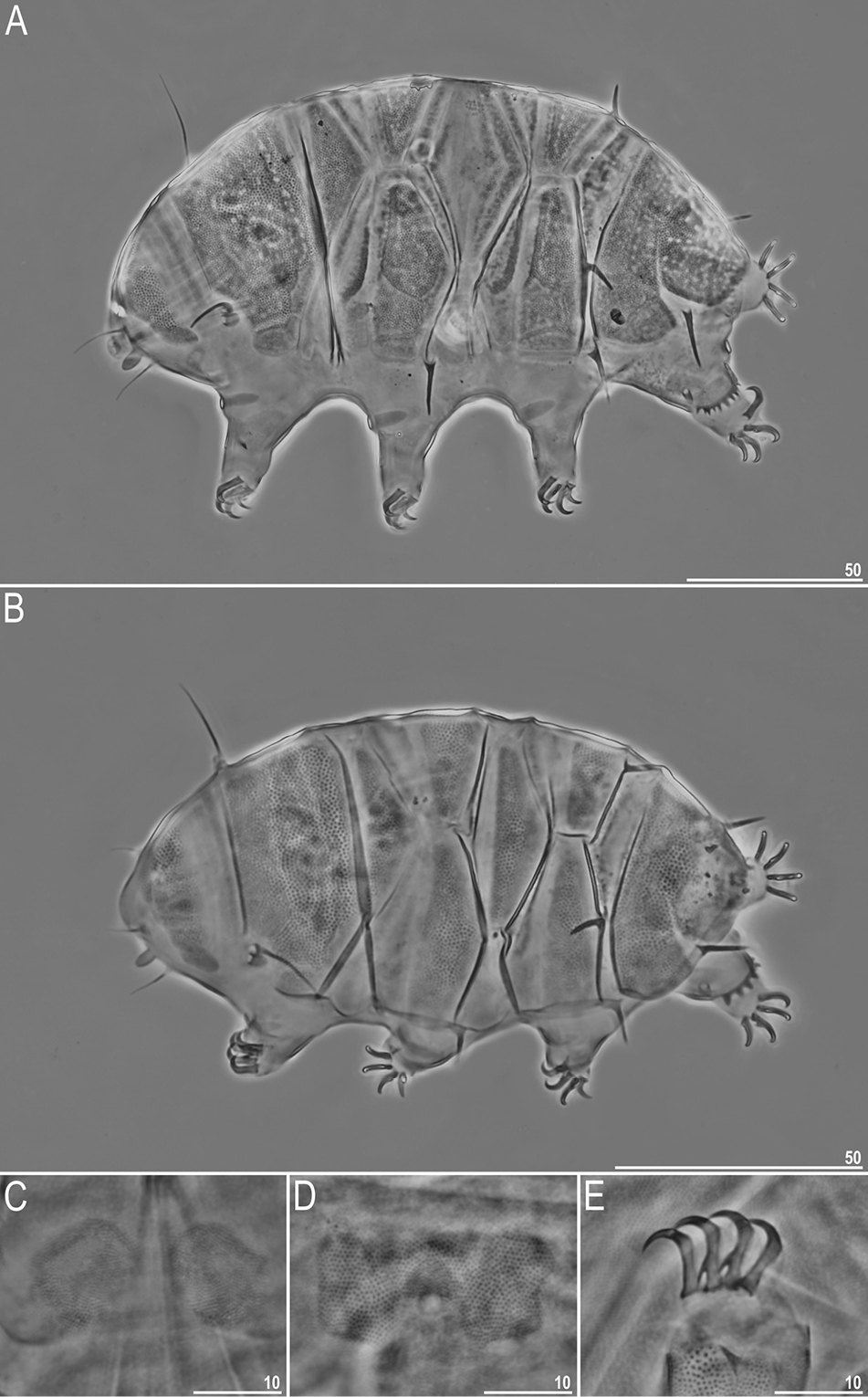
Peinaleopolynoe orphanae (A), Peinaleopolynoe elvisi (B), Peinaleopolynoe goffrediae (C) and Peinaleopolynoe mineoi (D) are four new scale worms from the Pacific Ocean. Credits to Hatch et al. (2020).*
A new sponge-associated starfish was named Astrolirus patricki and I think the reason for that name is evident enough, right? Credits to Zhang et al. (2020).*
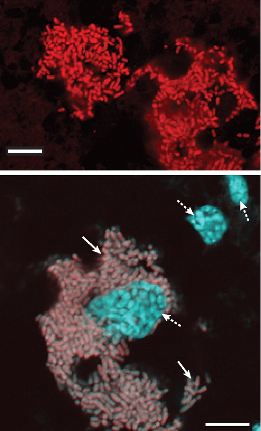
Bacteria are found almost everywhere across our planet and they are essential for the survival of every other lifeform, including the fascinating, and for some disgusting, cockroaches. One special cockroach-friendly genus of bacteria has the adequate name Blattabacterium, whose best-known species is Blattabacterium cuenoti, which I decided to call the “common cockroach bacterium”.
This interesting species, like all species of Blattabacterium, is an obligate endosymbiont of cockroaches, meaning that it can only exists inside cockroach cells. More specifically, the common cockroach bacterium lives inside the cells of the fat bodies of cockroaches, i.e., their adipose tissue. It was found living inside all cockroach species examined to date with the exception of the genus Nocticola. It is also found inside the termite Mastotermes darwinensis because, if you did not know yet, termites are nothing more than highly specialized cockroaches. Thus, it is thought that this bacterium first “infected” the ancestor of all modern cockroaches about 140 million years ago and has only been lost in two lineages, the one from Nocticola and the one from termites.
Blattabacterium cuenoti cells shown in red (above) and gray (below). The cyan areas in the bottom image represent the nucleus of the cockroach cells. Extracted from Sabree et al. (2009).
Although many cockroaches are generalist feeders, being able to feed on almost everything, the main diet of all species is decaying plant material, and this is a relatively nitrogen-poor food. In order to increase their nitrogen intake, cockroaches store uric acid, a common product of protein metabolism. Most animals, including humans, excrete uric acid in their urine, but cockroaches store it in their adipose tissue. Thus, it was originally thought that the cockroach bacteria, by living close to uric acid reserves in the adipose tissue, could use uric acid directly as a food source, but studies have found this is not the case.
When necessary, cockroaches release this uric acid and it is broken down into urea or ammonia by bacteria living in their guts. After that, the common cockroach bacteria can use those compounds to synthesize glutamate, essential amino acids and vitamins for the cockroach.
Since they cannot use uric acid directly, it is a mystery why the common cockroach bacteria lives so close to the place where this substance is stored. One suggestion is that it was originally able to use uric acid but lost this ability by genome reduction.
The functional gene categories of Blattobacterium are very similar to those of Blochmannia, an endosymbiotic bacterium from carpenter ants, which also feed on plant material. However, Blochmannia is very distantly related to Blattobacterium, suggesting that their similar genomes are the result of convergent evolution caused by similar lifestyles.
When something works, nature invents it more than once.
López-Sanchez MJ, Neef A, Peretó J, Patiño-Navarrete R, Pignatelli M, Latorre A, Moya A (2009) Evolutionary Convergence and Nitrogen Metabolism in Blattabacterium strain Bge, Primary Endosymbiont of the Cockroach Blattella germanica. PLoS Genetics 5(11): e1000721. 10.1371/journal.pgen.1000721
Patiño-Navarrete R, Moya A, Latorre A, Peretó J (2013) Comparative Genomics of Blattabacterium cuenoti: The Frozen Legacy of an Ancient Endosymbiont Genome. Genome Biology and Evolution 5(2): 351–361. https://doi.org/10.1093/gbe/evt011
Sabree ZL, Kambhapati S, Moran NA (2009) Nitrogen recycling and nutritional provisioning by Blattabacterium, the cockroach endosymbiont. PNAS 106(46): 19521–19256. https://doi.org/10.1073/pnas.0907504106
Flatworms are the fourth largest animal phylum after arthropods, mollusks and chordates and most species known to date belong to the clade Neodermata, which includes parasitic species such as flukes and tapeworms, some of which infect humans. Among tapeworms, the species that infect humans and belong to the genus Taenia are certainly the most popular, but it is expected that all vertebrates can have at least one tapeworm parasite, so that it is only a matter of time and opportunity for us to discover them all.
Among the species known to parasitize horses, the most widespread is Anoplocephala perfoliata, which I decided to call the common horse tapeworm. As tapeworms in general, the adult common horse tapeworm lives in the intestine of its definitive host, in this case a horse.
Different from species of Taenia, which can grow up to several meters in length, species of Anoplocephala are much smaller. The whole body of adult specimens measures about 5 to 8 cm in length and 1 to 2 cm in width and is divided into the same parts seen in other tapeworms. The anteriormost part of the body includes the scolex, which has 4 large suckers. Although the scolex of most tapeworms measures less than 1 mm, in the common horse tapeworm it reaches up to 3 mm.
A preserved specimen. Photo extracted from alchetron.com
After the scolex there is a small neck of undifferentiated tissue that grows constantly to build new proglottids, which form the rest of the body. Proglottids are connected to each other in a chain fashion and the posteriormost ones are continuouly lost and released into the environment. Each proglotid contains male and female sexual organs and is released when it contains mature eggs.
Mature proglottids are released in the environment through the horse’s feces and release their eggs on the ground and the vegetation. The eggs can survive outside a host for as long as 9 months. During this time, they hope to be accidentally ingested by oribatid mites that live in pastures. If this happens, the egg hatches inside the mite due to the mechanical action of the mite’s mouth parts and releases the first-stage larva called the oncosphere.
An egg of Anoplocephala perfoliata. The small 20-µm-diamter sphere is the oncosphere waiting to be released. Photo by Martin K. Nielsen, extracted from msdvetmanual.com
When the oncosphere reaches the mite’s gut, it is activated, probably via ions present in this environment, and uses a group of hooks to penetrate the mite’s tissues. After about 8 to 20 weeks, the oncosphere develops into a cysticercoid. This stage looks like an inverted miniaturized tapeworm inside a bladder-like vesicle, having already a protoscolex inside it.
While horses are grazing, they always ingest some invertebrates together with the grass. It they happen to ingest an infected mite, the cysticercoids are released during digestion, evert the protoscolex and attach to the intestinal walls of the horse. There, the tapeworm develops into an adult, restarting the cycle.
Attached to the intestine of their hosts, tapeworms do not feed on blood or other tissues as many parasites do. Instead of that, they collect nutrients directly from the host’s gut by absorbing them via the worm’s body surface.
For a long time its has been thought that the common horse tapeworm was a harmless parasite since most horses did not seem to have any symptom and the tapeworms were often only discovered during dissection after the horse’s death by other causes. The preferred area for the common horse tapeworm to attach is the caecum and the ileocaecal junction but in heavily infected animals some individuals can be found in suboptimal sites throughout the small and large intestines. In such heavily infected horses, the tapeworms can cause colics and even intestinal obstruction.
Large number of adult tapeworms in a heavily infected horse. Credits to Tomczuk et al. (2014).*
The common horse tapeworm can infect other equids as well, such as donkeys and zebras. Ironically, domesticated horses seem to be the most infected individuals exactly because horse owners treat them with anthelmintics. Most modern anthelmintics do not affect tapeworms and only remove other parasites, such as roundworms, which reduces competition and allows tapeworms to thrive.
Gasser RB, Williamson RMC, Beveridge I (2005) Anoplocephala perfoliata of horses – significant scope for further research, improved diagnosis and control. Parasitology 131(1): 1–13. https://doi.org/10.1017/S0031182004007127
Tomczuk K, Kostro K, Szczepaniak KO, Grzybek M, Studzińska M, Demkowska-Kutrzepa M, Roczeń-Karczmarz M (2014) Comparison of the sensitivity of coprological methods in detecting Anoplocephala perfoliata invasions. Parasitology Research 113(6): 2401–2406. doi: 10.1007/s00436-014-3919-4
Two mosquitoes of the genus Aedes, Aedes aegypti and Aedes albopictus, are invasive species in tropical and subtropical regions worldwide. While A. aegypti is native from Africa, A. albopictus is originally from southeast Asia, but both species have been spread by humans and continue to increase their range.
Both species are known as vectors of several diseases that affect humans, especially those caused by Flavoviruses, which include the Yellow fever, Dengue fever and Zika fever. Chikungunya, caused by a species of Alphavirus is also transmitted to humans by them. Moreover, they can also transmit some nematodes, such as the heartworm that infects the heart of dogs and other carnivores.
Aedes aegypti biting a human and having a delicious bloody meal. Photo by James Gathany.
Because A. aegypti and A. albopictus pose such a huge threat to public health, getting rid of them is top priority. Here in Brazil, there is a massive national campaign to reduce the ability of Aedes to reproduce by avoiding containers with still water in the open, such as flower vases, buckets, uncovered barrels, discarded tires and virtually everything that can retain water long enough for the larvae to develop. I have to say, though, that this all seems to be useless. The mosquitoes continue to spread and the cases of dengue fever continue to grow. The fact is that the mosquitoes will find a place to lay their eggs. If they don’t find it in your backyard, they will find it in the forest or any vacant lot.
Instead of forcing them to lay their eggs where we cannot see, we should stimulate them to lay their eggs around us and then kill the larvae. Several aquatic predators have been tested as potential allies, including larvivorous fish, dragonfly nymphs, copepods, planarians and even other mosquitoes whoses larvae eat the larvae of Aedes! The use of these predators showed mixed results. Larvivorous fish are difficult to maintain in water tanks at home and dragonfly nymphs are too generalist as predators.
Now a new predator has been suggested: a plant! Yes, a carnivorous plant of the genus Utricularia, which includes species known as bladderworts. These aquatic plants have little bladder-like structures that function as traps to capture small animals. The bladder is hollow and has an internal negative pressure in relation to the environment surrounding it. This negative pressure is created by water being constanly pumped out of the bladder through its walls via active transport. The bladder’s opening is covered by a small lid that avoids water to fill it again when the trap is set. Surrounding the lid, there is a group of bristle-like protuberances. When an animal is moving through the water and moves one of those bristles, they slightly deform the lid, breaking the seal and allowing water to enter the bladder. The negative pressure then sucks water quickly into the bladder, dragging the small animal with it. Then it is only a matter of time for the poor animal to be digested.
Watch the plant in action.
A group of researchers at the University of Rhode Island, USA, tested whether Utricularia macrorhiza, the common North American bladderworth, could be an effective predator of mosquito larvae. By adding U. macrorhiza to containers with larvae of A. aegypti and A. albopictus, they were able to kill 95 to 100% of the larvae in only five days. That’s an amazing result, don’t you think?
Bladderwort with several Aedes larvae (marked with asterisks) in its traps. Credits to Couret et al. (2020).*
Since bladderworts are much easier to raise in tanks and other containers in your backyard than animal predators such as fish and dragonflies, they are a promising new alternative to control the populations of this disease-carrying insects.
So, are you eager to raise some aquatic carnivorous plants to help fight these heinous mosquitoes?


 This work is licensed under a Creative Commons Attribution-NonCommercial-NoDerivs 4.0 International License.
 This work is licensed under a Creative Commons Attribution-NonCommercial-ShareAlike 4.0 International License.