Quantcast
Channel: Earthling Nature
Viewing all 556 articles
Browse latest View live

Friday Fellow: Tulip Tree

$
0
0

by Piter Kehoma Boll

When people reach a new locality and find new species, they have to think of a way to name them, which can happen by borrowing a name from a local language or make up a new name from one’s own language. When the first Europeans reached North America, they discovered a beautiful tree growing in what is now eastern United States. The Miami people called it oonseentia, but we all know how Europeans treated native Americans back then. So, instead of borrowing this word, they made up a new and completely misleading name: tulip tree.

Cultivated tulip tree in Belgium. Photo by Jean-Pol Grandmont,**

Linnaeus gave this tree its currently accepted binomial name: Liriodendron tulipifera, literally meaning “lily tree that carries tulips”. However, this species has nothing to do with lilies and tulips, being actually closely related to magnolias.

Reaching up to 50 m in height, and rarely becoming even taller, the tulip tree has a brown and furrowed bark and smooth and lustrous branches. The leaves have four large lobes that, if you make a lot of effort, may look a little bit like a violin, which makes it have an additional common name: fiddletree.

Typical “violin”-shaped leaf from a tree in Virginia, USA. Credits to Wikimedia user PumpkinSky.*

The flowers of the tulip tree appear in summer and very superficially resemble a tulip, although their structure is quite different. They have three green sepals and six petals that are arranged in a spiral that continues inward to form the stamens and then the pistils, which form a central cone. This arrangement is considered primitive within angiosperms and kind of look as something between a gymnosperm cone and a true angiosperm flower.

Flowers on a tree in New Jersey, USA. Photo by Wikimedia user Famartin.*

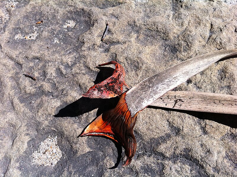
The mature seeds, called samaras, are dispersed by wind. They develop in autumn and are stored in a type of cone-like fruit. As a typical temperate species, the tulip tree is deciuous, shedding its leaves in winter.

Frosted fruits in winter in Virginia, USA. Photo by Jörg Peter.

The tulip tree is considered a species that dominates the first century of a forest since its establishment. It is a shade-intolerant species, so when other trees start to grow among them and block much of the sunlight, they tend to perish.

Due to its beauty, the tulip tree has become an ornamental plant and several cultivars have been developed. Its wood is also used for construction, and Native Americans used to build canoes from its trunks. Due to its wood, the tulip tree has also received the common name “yellow poplar” although it is not closely related to the true poplars, such as the black and white poplar. In fact, their wood is not that similar, with the tulip tree or “yellow poplar” wood being of much higher quality. In other words, the name “yellow poplar” is as misleading as the name “tulip tree”.

Big and old tulip trees in the Joyce Kilmer Memorial Forest, North Carolina, USA. Photo by Wikimedia user Notneb82 .**

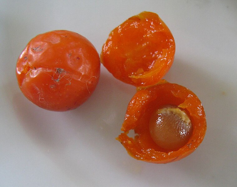
The orange part of the petals contain nectar that, when collected by bees, creates a special and strong honey that is usually considered unsuitable for table honey but highly regarded by bakers.

Native Americans and early European settlers used the tulip tree to treat malaria, and modern studies have confirmed that some of its constituents show antiplasmodial activity, as well as antioxidant, antimicrobial and cytotoxic properties, having the potential to help the development of new antibiotics and anticancer drugs.

– – –

Like us on Facebook!

Follow us on Twitter!

– – –

References:

Quassinti L, Maggi F, Ortolani F, Lupidi G, Petrelli D, Vitali LA, Miano A, Bramucci M (2019) Exploring new applications of tulip tree (Liriodendron tulipifera L.): leaf essential oil as apoptotic agent for human glioblastoma. Environmental Science and Pollution Research 26:30485–30497. https://doi.org/10.1007/s11356-019-06217-4

Wikipedia. Liriodendron tulipifera. Available at: <https://en.wikipedia.org/wiki/Liriodendron_tulipifera>. Access on June 18, 2020.

– – –

*Creative Commons License This work is licensed under a Creative Commons Attribution-ShareAlike 4.0 International License.

**Creative Commons License This work is licensed under a Creative Commons Attribution-ShareAlike 3.0 Unported License.


Friday Fellow: Peach Leaf-Curl Fungus

$
0
0

by Piter Kehoma Boll

Fungi are essential for the maintenance of life on Earth, but some of them are also a pain in the leaf, such as Taphrina deformans, which causes the disease known as peach leaf curl.

As you can imagine, peach leaf curl is a disease that affects peach trees (as well as nectarines, which are nothing but a variety of peach) and eventually can occur on almond trees too. The hyphae grow between the leaf cells and secrete enzymes that degrade cellulose, as well as indole-3-acetic acid, a type of auxin, i.e., a plant hormone that induces cell growth and division. As a result, infected leaves start to curl inward and downward and turn from green to pale yellow and eventually red.

Taphrina deformans causing leaf curl on a peach tree in Portugal. Credits to Duarte Frade.*

When the peach leaf-curl fungus is mature, it produces vertical hyphae that grow toward the surface of the leaf, spread just below the cuticle, and form asci, sac-like cells filled by ascospores, the sexual spores. The asci break through the leaf’s surface and cause a whitish aspect. The ascospores produce conidia, the asexual spores, and those are released in the environment, where they wait for the ideal conditions to germinate.

Conidia often remain attached to the branches of the tree and grow in a yeast-like fashion. They infect new leaves as soon as they start to grow. In order to germinate and infect leaves, conidia require about 3 mm of rainfall followed by 12 days with enough humidity and temperatures below 19 °C. As a result, infections are much more common in temperate regions and do not occur every year, as sometimes the requirements are not met. Fungicide is often efficient to stop the infections, but if humidity is too high and the fungus has spread too much, the treatment may not be efficient enough.

A very curly leaf with the whitish surface cased by the asci. Photo by Jerzy Opioła.**

Infected leaves fail to make photosynthesis effectively and die earlier. As a result, the plant becomes weak and produces few or no fruits, which may cause total yield loss.

The genome of the peach leaf-curl fungus has been sequenced and showed to be considerably small compared to other fungal pathogens. Nevertheless, about 5% of its genes are only found in other fungal pathogens, including, for example, enzymes that are able to break the cuticle of plants, which is necessary for infection to occur.

Genes capable of producing plant hormones, such as the auxin mentioned above, appear to be absent in closely related species. Although the idea that they may have been acquired from the plants themselves via horizontal gene transfer has been raised, a deeper analysis suggest that they are formed by very different pathways and probably evolved independently.

When something works, nature invents it more than once, although sometimes the second invention serves as a way to cheat the first one.

– – –

Like us on Facebook!

Follow us on Twitter!

– – –

References:

Cissé OH, Almeida JMGCF, Fonseca A, Kumar AA, Salojärvi J, Overmyer K, Hauser PM, Pagni M (2013) Genome Sequencing of the Plant Pathogen Taphrina deformans, the Causal Agent of Peach Leaf Curl. mBio 4(3):e00055-13. https://doi.org/10.1128/mBio.00055-13

Martin EM (1940) The morphology and cytology of Taphrina deformans. American Journal of Botany 27(9):743–751. https://doi.org/10.2307/2436901

Wikipedia. Taphrina deformans. Available at <https://en.wikipedia.org/wiki/Taphrina_deformans>. Access on 25 June 2020.

– – –

*Creative Commons License This work is licensed under a Creative Commons Attribution 4.0 International License.

**Creative Commons License This work is licensed under a Creative Commons Attribution-ShareAlike 4.0 International License.

New Species: June 2020

$
0
0

by Piter Kehoma Boll

Here is a list of species described this month. It certainly does not include all described species. You can see the list of Journals used in the survey of new species here.

Terrilactibacillus tamarindi is a new lactic acid bacteria isolated from the bark of tamarind trees in Thailand. Credits to Kingkaew et al. (2020).*
Phytoactinopolyspora mesophila is a new actinobacterium isolated from a saline-alkaline soil in China. Credits to Feng et al. (2020).*

Bacteria

Cupriavidus agavae is a new proteobacterium isolated from the rhizosphere of agave plants in Mexico. Credits to Arroyo-Herrera et al. (2020).*

Archaeans

Halobacterium bonnevillei (a), Halobaculum saliterrae (b) and Halovenus carboxidivorans (c) are three new archaeans from saline crusts and soils. Credits to Myers and King (2020).*

SARs

Actinostachys minuta is a new grass fern from the Philippines, Credits to Amoroso et al. (2020).*

Plants

Argyreia pseudosolanum is a convolvulacean from Thailand whose flowers resemble a species of Solanum. Credits to Traiperm & Suddee (2020).*
Senecio festucoides is a new composite from Chile. Credits to Calvo & Moreira-Muñoz (2020).*

Amoebozoans

Curvularia paraverruculosa is a new Pleosporalean isolated from soil samples in Mexico. Credits to Iturrieta-González et al. (2020).*

Fungi

Hygrophorus fuscopapillatus is a new mushroom from Southern China. Credits to Wang et al. (2020).*

Poriferans

Cnidarians

Flatworms

Dugesia umbonata is a new planarian from China. Credits to Song et al. (2020).*

Mollusks

Annelids

Bryozoans

Nematomorphs

Gordius chiashanus is a new millipede-parasitizing horsehair worm from Taiwan. Credits to Chiu et al. (2020).*

Nematodes

Chelicerates

Myriapods

Plusioglyphiulus biserratus (top) and Plusioglyphiulus khmer (bottom) are two new millipedes from Cambodia. Credits to Likhitrakarn et al. (2020).*
Fredius ibiapaba is a new freshwater crab from northeastern Brazil. Credits to Chávez et al. (2020).*

Crustaceans

Cycladiacampa irakleiae is a new cave-dwelling dipluran from Irakleia Island, Cyclades Islands. Credits to Sendra et al. (2020).*
Tachycines trapezialis is a new cave cricket from China. Credits to Zhou & Yang (2020) .*

Hexapods

Dolichomitus mariajosae is a new parasitoid wasp from Colombia, Credits to Araujo et al. (2020).*
Oromia orahan is a new subterranean beetle from La Gomera, Canary Islands. Credits to García et al. (2020).*

Echinoderms

Chondrichthyans

Actinopterygians

Plectranthias hinano is a new perchlet from the Pacific. Credits to Shepherd et al. (2020).*

Amphibians

Dendropsophus bilobatus is a new tree frog from the Amazon Forest in Brazil. Credits to Ferrão et al. (2020).*
Platypelis laetus is a new narrow-mouthed frog from Madagascar. Credits to Rakotoarison et al. (2020).*

Reptiles

– – –

Like us on Facebook!

Follow us on Twitter!

– – –

*Creative Commons License This work is licensed under a Creative Commons Attribution 4.0 International License.

Friday Fellow: Brown’s Dagger Nematode

$
0
0

by Piter Kehoma Boll

Nematodes are famous because of their parasitic members, which do not only parasitize animals but also plants. People that deal with gardening or agriculture may know that sometimes a plant becomes sick because of “nematodes”.

A genus of nematodes that is commonly associated with grapevines is Xiphinema, whose species are known as dagger nematodes. The two most widely studied species are Xiphinema americanum, the American dagger nematode, and Xiphinema index, the California dagger nematode, but during the last decades it became clear that those species are actually a complex of very similar species and new ones are constantly been described. One of them, described in 2016, is Xiphinema browni, which I decided to call Brown’s dagger nematode. It was named after the nematologist Derek J. F. Brown.

Brown’s dagger nematode was found associated with the roots of grapevines and apple trees in Slovakia and the Czech Republic. Among 86 identified females there was only one male, indicating a huge disparity in sex ratios and the probability that females are parthenogenetic, i.e., they can lay fertile eggs without being fertilized by a male. Females measure up to 2.5 mm in length and the only known male measured 1.8 mm.

Female (left) and male (right) of Xiphinema browni. Modified from Lazarova et al. (2020).*

Since Brown’s dagger nematode was found associated with grapevines, its life cycle is likely similar to that of most other dagger nematodes. Adults are external parasites of grapevine roots and eventually of other woody plants. They live on the root surface and use their long odontostyles (a needle-like proboscis) to perforate the roots and suck the content of their vascular tissue. As a reaction, the plant produces swollen club-like galls on the root tips. The root then branches behind the swollen tip, only to be attacked again, developing another gall and having to branch again. This starts to weaken the plant, which can compromise grape production.

Anterior end of a female with the odontostyle slightly exposed. Modified from Lazarova et al. (2020).*

Females lay their eggs scattered through the soil, not forming clusters, and juveniles pass through about 4 stages in the soil before turning to the parasitic mode.

As another grapevine-feeding dagger nematode, Brown’s dagger nematode is probably also a vector of the grapevine fanleaf virus, which is transmitted to grapevines by the California dagger nematode. This happens when the nematode feeds on an infected plant and then moves to a healthy plant, carrying the virus with it. Grapevine fanleaf causes chlorosis (loss of chlorophyll) and distorts the leaves, making them look like fans, hence the name. As you can imagine, the poor plant becomes even weaker than it already was due to the nematodes sucking it. This can be a nightmare to vineyard owners.

The grapevine fanleaf virus can be a devastating disease for grapevines but in the nematode’s body it seems to have benefitial effects, increasing the survival of these small roundworms. Perhaps this stimulates the dagger nematodes to spread it further, in a sort of “evil coalition”.

– – –

You may also like:

Tospovirus and thrips: an alliance that terrifies plants

– – –

Like us on Facebook!

Follow us on Twitter!

– – –

References:

Lazarova S, Peneva V, Kumari S (2016) Morphological and molecular characterisation, and phylogenetic position of X. browni sp. n., X. penevi sp. n. and two known species of Xiphinema americanum-group (Nematoda, Longidoridae). ZooKeys 574:1–42. https://doi.org/10.3897/zookeys.574.8037

van Zyl S, Vivier MA, Walker MA (2012) Xiphinema index and its Relationship to Grapevines: A review. South African Journal of Enology and Viticulture 33(1):21–32.

Wikipedia. Xiphinema. Available at <https://en.wikipedia.org/wiki/Xiphinema>. Access on 29 June 2020.

– – –

*Creative Commons License This work is licensed under a Creative Commons Attribution 4.0 International License.

Friday Fellow: Red Bogmoss

$
0
0

by Piter Kehoma Boll

Among the many different ecosystems found on Earth, bogs are particularly interesting. These peculiar wetlands are basically a large amount of water-soaked plant matter, either dead or alive. Usually very acidic, bogs have very low decomposition rates, so plant matter tends to accumulate more and more, sometimes reaching several meters in depth.

The main organisms responsible for the formation of bogs are mosses of the genus Sphagnum, commonly known as bogmosses or peatmosses (peat being the plant material that forms the bogs). Found all around the world, bogmosses have the ability to absorb huge amounts of water, just like a sponge, and in dry conditions they can release this water into the surrounding areas, helping them stay humid.

Red bogmoss in Canada. Credits to iNaturalist user maddieology.*

One bogmoss species, the red bogmoss, Sphagnum capillifolium, is found in the northern half of North America and Europe, being an important and genetically diverse species. In fact, it is likely that the red bogmoss is actually a complex of many very similar species. Its scientific name, capillifolium, meaning “hair-leaf”, refers to the peculiar shape of the plant, which grows in straight and densely packed branches that bent outwards at the top, resembling tresses.

Greener specimens in the USA. Photo by Joe Walewski.*

Although most bogmoss species are green like any regular plant, the red bogmoss and closely related species can have a reddish color. However, this color is not caused by pigments in their plastids but by a pigment, sphagnorubin, found in their cell walls. The presence or not of sphagnorubin seems to be determined by certain combinations of temperature, light and hormones. The exact function of sphagnorubin is unknown, but there have been suggestions that it may help protect the plant from herbivory. It i also possible that this reddish color works as a sunscreen, protecting the plant’s chloroplasts from intense radiation since sphagnorubin absorbs UV and blue light.

A very red and water-soaked mass in Scotland. Credits to Andrew Melton.*

Bogmosses in general are not attractive to herbivores because they contain high amounts of phenolic compounds, such as tannins, which gave them an adstringent and bitter taste. These phenolic compounds are also the main reason why peat takes such a long time to decompose. As a result, bogs function as huge carbon reservoirs, and about 10 to 15% of all carbon stock on the planet is in the form of Sphagnum. In fact, the amount of carbon fixed by all other photosynthetic lifeforms on Earth every year is lower than the amount held in bogs.

Some slightly red ones in England. Photo by Jeremy Barker*.

Sphagnum is, thus, an essential genus to keep the levels of carbon dioxide in the atmosphere low and the red bogmoss is even more important because it seems to be a very tolerant species that can survive in both shaded and sunny environments, as well as conditions with low and high levels of nitrogen and may, therefore, resist human interference better than other bogmosses.

– – –

Like us on Facebook!

Follow us on Twitter!

– – –

References:

Bonnett SAF, Ostle N, Freeman C (2009) Short-term effect of deep shade and enhanced nitrogen supply on Sphagnum capillifolium morphophysiology. Plant Ecology 207: 347–358. https://doi.org/10.1007/s11258-009-9678-0

Gerdol R, Bonora A, Marchesini R, Gualandri R, Pancaldi S (1998) Growth Response of Sphagnum capillifolium to Nighttime Temperature and Nutrient Level: Mechanisms and Implications for Global Change. Arctic and Alpine Research 30(4): 288–395. https://doi.org/10.1080/00040851.1998.12002914

Verhoeven JTA, Liefveld WM (1997) The ecological significance of organochemical compounds in Sphagnum. Acta botanica neerlandica 46(2): 117–130. http://natuurtijdschriften.nl/record/541086

– – –

*Creative Commons License This work is licensed under a Creative Commons Attribution-NonCommercial 4.0 International License.

Friday Fellow: Tiger Leech

$
0
0

by Piter Kehoma Boll

I never had the opportunity to visit Borneo, but people say that, when you walk inside the rainforest there, it is impossible not to meet today’s “adorable” fellow, the so-called tiger leech, Haemadipsa picta.

As you may know, many leeches feed on blood, and the tiger leech is no exception. Its genus name, Haemadipsa, even means “blood thirst”. However, different from most leeches, which are aquatic, the tiger leech and many of its close relative live on land or, in the case of the tiger leech, most often on vegetation in Southeast Asia.

A tiger leech preparing to attack a passing mammal, most likely the human taking its photo, which in this case would be Kristof Zyskowski.*

For a leech, the tiger leech is very beautiful. Its dorsum has a series of black, yellow and brown longitudinal stripes and bands which often acquire a green tinge toward the anterior end. The posterior end has a large sucker, which sometimes has a green color too, and the tiger leech use it to remain attached to the substrate, usually vegetation.

The food of the tiger leech consists mainly of the blood of mammals that happen to walk across its path. They detect their prey by a combination of visual, mechanical and chemical cues. They can see the prey with a series of small eyes, feel them with mechanoreceptors and smell the carbon dioxide that they exhale while breathing.

Sometimes the prey makes fun of the predator. Credits to Ellen Rykers.**

The tiger leech “knows” where it is more likely to find a passing mammal: trails inside the forest. Thus, they gather on the leaves and branches of plants growing along such paths. Smaller, younger specimens, prefer to stay closer to the ground to avoid spending too much energy climbing up, while adult specimens (which reach up to 33 mm in length) wait for their meal at a height of about 1.5 m, rarely climbing up to 2 m. Considering that the largest non-human mammals to walk across the forest do not get higher than 1.7 m, they don’t need to climb above that limit.

A thirsty tiger leech in Taiwan. Photo by Liu JimFood.**

When a juicy mammals is passing through the trail, the leeches, which are eagerly waiting with their bodies stretched forward, jump on the animal’s body, cut their skin with their sharp mouth parts, and start to drink their delicious blood. It is said that the bite of the tiger leech is very painful, different from most leeches, and it is hard to make the wound stop bleeding, probably because they release a considerable amount of anticoagulant. Not a pleasant experience, I guess.

– – –

Like us on Facebook!

Follow us on Twitter!

– – –

References:

Gasiorek P, Różycka H (2017) Feeding strategies and competition between terrestrial Haemadipsas leeches (Euhirudinea: Arhynchobdellida) in Danum Valley rainforest (Borneo, Sabah). Folia Parasitologica, 64: 031. https://doi.org/10.14411/fp.2017.031

Kendall A (2012) The effect of rainforest modification on two species of South-East Asian terrestrial leeches, Haemadipsa zeylanica and Haemadipsa picta. Master Thesis.

– – –

*Creative Commons License This work is licensed under a Creative Commons Attribution-NonCommercial-NoDerivs 4.0 International License.

**Creative Commons License This work is licensed under a Creative Commons Attribution-NonCommercial 4.0 International License.

Friday Fellow: Busy Lizzie

$
0
0

by Piter Kehoma Boll

You probably know today’s fellow. You may have this plant in your garden or have seen it in someone else’s garden, in parks or, if you live in tropical and subtropical areas, everywhere along roads or inside the forest. Scientifically known as Impatiens walleriana, it is commonly called busy Lizzie, sultana, impatiens or balsam, although impatiens and balsam can be used for other species of the genus Impatiens.

Busy Lizzie in Vietnam. Credits to Wikimedia user Prenn.**

This lovely plant has tender and succulent stem with a light green to dark red color that becomes semiwoody at the base. The leaves are tender, ovate, smooth and have a serrated border. The base of the leaves have small extrafloral nectaries. The flowers have 5 petals and 5 sepals and are bilaterally symmetric, with the lower sepal forming a long spur containing nectar. The fruits develops into a small capsule that explodes as a strategy to spread the seeds.

Extrafloral nectaries dripping. Photo by Wikimedia user Mariluna.**

The busy Lizzie is native from eastern Africa and is cultivated worlwide as an ornamental plant, with several different cultivars. The flowers can be white, pink, salmon, red, magenta, purple and many other varieties, sometimes even with more than one color. Interesting, though, a study revealed that white is the favorite color for butterflies that visit the plant, while red is the least favorite color. In fact, although different butterfly species show slightly different preferences for color, no species seems to like visiting red flowers.

A specimen growing in South Africa. Photo by Stuart Billingham.*

After being transported to other continents, the busy Lizzie become naturalized in many regions, especially tropical and subtropical areas in the Americas and southeast Asia. The impact of its spread throughout these areas is not clear as far as I know.

Several varieties being cultivated in Bangalore, India. Photo by Ramesh Ng.**

One interesting fact is that mosquitoes seem to love feeding on its extrafloral nectaries, and studies have shown that they are the preferred nectar source for mosquitoes of the genus Aedes. Thus, the busy Lizzie is know being studied as a potential way to control the populations of these disease-carrying insects by the development of genetically modified varieties with inseticidal nectar.

Other studies have shown that the busy Lizzie is a cadmium accumulator, i.e., it can remove large amounts of cadmium from the soil and has the potential do be used as a tool for decontamination. Busy Lizzie is very busy, indeed.

– – –

Like us on Facebook!

Follow us on Twitter!

– – –

References:

Lai H-Y (2015) Subcellular distribution and chemical forms of cadmium in Impatiens walleriana in relation to its phytoextraction potential. Chemosphere 138:370–376. https://doi.org/10.1016/j.chemosphere.2015.06.047

Lim TK (2013). Impatiens walleriana. Edible Medicinal and Non-Medicinal Plants: 548–550. https://doi.org/10.1007/978-94-007-7395-0_34

Mandle M, Warren DL, Hoffmann MH, Petersen AT, Schmitt J, von Wettberg EJ (2010) Conclusions about Niche Expansion in Introduced Impatiens walleriana Populations Depend on Method of Analysis. PLoS ONE 5(12): e15297. https://doi.org/10.1371/journal.pone.0015297

Morris AB (2005) Functional differences among color morphs of Impatiens walleriana (Balsaminaceae).

Pruett G, Hawes J, Varnado W, Deerman H, Goddard J, Burkett-Cadena N, Kearney C (2020) The readily transformable Impatiens walleriana efficiently attracts nectar feeding with Aedes and Culex mosquitoes in simulated outdoor garden settings in Mississippi and Florida. Acta Tropica. https://doi.org/10.1016/j.actatropica.2020.105624

– – –

*Creative Commons License This work is licensed under a Creative Commons Attribution-NonCommercial 4.0 International License.

**Creative Commons License This work is licensed under a Creative Commons Attribution-ShareAlike 3.0 Unported License.

Friday Fellow: Pineapple Sea Cucumber

$
0
0

by Piter Kehoma Boll

Seafood is often a highly regarded food all around the world and include all sorts of marine organisms that humans found out that are edible. In Southeast Asia, one of these delicacies is a sea cucumber, Thelenota ananas, known as pineapple sea cucumber, tripang or prickly redfish.

A nice pineapple sea cucumber in the Maldivas. Photo by Albert Kang.*

The pineapple sea cucumber is an echinoderm, a group which also includes seastars, brittle stars, sea urchins, sand dollars and sea lilies. It is found in tropical and subtropical waters of the Indo-Pacific, occurring from the Red Sea southward along the east coast of Africa and eastward to Polynesia, being common in coral reefs, although in low densities.

Reaching up to 70 cm in length and 6 kg in weight, the pineapple sea cucumber is a relatively large sea cucumber. It has a reddish-orange and black color, usually brighter on the underside, and has many soft star-like projections (“teats”) all over the body.

A detail showing the star-shaped “teats”. Photo by Nick Hobgood.**

Like most sea cucumbers, the pineapple sea cucumber is a herbivore. As a larva it probably feeds on phytoplankton and, as an adult, on larger algae, including calcaerous green algae of the genus Halimeda. It grows slowly and has a long lifespan. In more subtropical areas, it reproduces in summer, from January to March, but in more tropical waters it is likely that it reproduces all year round. It can also involuntarily reproduce asexually if accidentally cut in half, with the anterior and posterior halves forming a new organism in a few weeks.

In the Northern Mariana Islands, with a human arm for comparison. Photo by John Starmer.*

As I said above, the pineapple sea cucumber is edible and is, in fact, a very healthy and promising food. As all sea cucumbers, it contains a fucoidan, a type of polysaccharide also found in brown algae and that has antioxidant and antiinflammatory properties. It is also rich in saponins, like other sea cucumbers and echinoderms, and these revealed to be good agents to reduce cholesterol levels and also have anticancer properties. More than that, the pineapple sea cucumber contains another compound, a glycosaminoglycan known as fucosylated chondroitin sulfate (FuCS-1), which revealed to have the ability to block HIV from entering cells and has, therefore, the potential to be explored for the development of new anti-HIV drugs, especially against some resistant variants.

A very red specimen in Malaysia. Photo by Tsu Soo Tan.*

Unfortunately, due to its slow development, the reproductive rate of the pineapple sea cucumber is unable to compensate its extraction from the ocean for human consumption. As a result, the natural populations have drastically decreased in the past decades, with a 60% reduction in New Caledonia and being almost extinct in some areas. As a result, it is listed as endangered in the IUCN’s red list. If we don’t start to respect this species by applying severe policies for harvesting it, we will end up losing a very precious fellow of our planet.

– – –

Like us on Facebook!

Follow us on Twitter!

– – –

References:

Conand C (1993) Reproductive biology of the holothurians from the major communities of the New Caledonian Lagoon. Marine Biology 116:439–450. https://doi.org/10.1007/BF00350061

Conand C, Gamboa R, Purcell S (2013) Thelenota ananasThe IUCN Red List of Threatened Species 2013: e.T180481A1636021. https://dx.doi.org/10.2305/IUCN.UK.2013-1.RLTS.T180481A1636021.en. Access on 30 July 2020.

Han Q, Li K, Dong X, Luo Y, Zhu B (2018) Function of Thelenota ananas saponin desulfated holothurin A in modulating cholesterol metabolism. Scientific Reports 8:9506. https://doi.org/10.1038/s41598-018-27932-x

Huang N, Wu M-Y, Zheng C-B, Zhu L, Zhao J-H, Zheng Y-T (2013) The depolymerized fucosylated chondroitin sulfate from sea cucumber potently inhibits HIV replication via interfering with virus entry. Carbohydrate Research 380:64–69. https://doi.org/10.1016/j.carres.2013.07.010

Reichenbach (1995) Potential for asexual propagation of several commercially important species of tropical sea cucumber (Echinodermata). Journal of the World Aquaculture Society 26(3):272–278. https://doi.org/10.1111/j.1749-7345.1995.tb00255.x

Wikipedia. Thelenota ananas. Available at < https://en.wikipedia.org/wiki/Thelenota_ananas >. Access on 30 July 2020.

Yu L, Xue C, Chang Y, Xu X, Ge L, Liu G, Wang Y (2014) Structure elucidation of fucoidan composed of a novel tetrafucose repeating unit from sea cucumber Thelenota ananas. Food Chemistry 146:113–119. https://doi.org/10.1016/j.foodchem.2013.09.033

– – –

*Creative Commons License This work is licensed under a Creative Commons Attribution-NonCommercial 4.0 International License.

**Creative Commons License This work is licensed under a Creative Commons Attribution-ShareAlike 3.0 Unported License.


New Species: July 2020

$
0
0

by Piter Kehoma Boll

Here is a list of species described this month. It certainly does not include all described species. You can see the list of Journals used in the survey of new species here.

Bacteria

Gordonia mangrovi is a new actinobacterium from mangrove soil in China. Credits to Xie et al. (2020).*
Craterlacuibacter sinensis is a new proteobacterium from a crater lake in China. Credits to Liu et al. (2020).*

SARs

Cryptists

Chondria tumulosa is a new red alga from Hawaii. Credits to Sherwood et al. (2020).*

Plants

Bredia hispida is a new melastome from China. Credits to Dai et al. (2020).*
Beautiful conidia of Spegazzinia musae, a new didymospheriacean fungus from Thailand. Credits to Samarakoon et al. (2020).*

Amoebozoans

Fungi

Sidera parallela is a new crust fungus from China. Photo by Bao-Kai Cui.*

Poriferans

Advhena magnifica is a new hexactinellid from the North Pacific. Credits to Castello-Branco et al. (2020).*

Cnidarians

Enteromyxum caesio is a new myxosporean parasitizing the fish Caesio cuning. Credits to Freeman et al. (2020).*

Rotiferans

Flatworms

Nemerteans

Eggs (A), juvenile (B) and adult (C) of Auriculella gagneorum, a new land snail from Hawaii. Credits to Yeung et al. (2020).*

Mollusks

Loimia borealis is a new terebellid from Chinese waters. Credits to Wang et al. (2020).*

Annelids

Bryozoans

Nematomorphs

Nematodes

Tardigrades

Chelicerates

Myriapods

Crustaceans

Heterochelamon huidongense is a new freshwater crab from southern China. Credits to Wang et al. (2020).*

Hexapods

Atrococcus rushuiensis is a new scale insect from China. Credits to Zhang et al. (2020).*
Teleopsis neglecta is a new stalk-eyed fly from Sri Lanka. Photo by Amila P Sumanapala.**

Echinoderms

Tunicates

Chondrichthyans

Lucifuga gibarensis is a new cave fish from Cuba. Credits to Hernández et al. (2020).*

Actinopterygians

Amphibians

Mammals

Lycodon cathaya is a new snake from China. Credits to Wang et al. (2020).*

Reptiles

– – –

*Creative Commons License This work is licensed under a Creative Commons Attribution 4.0 International License.

**Creative Commons License This work is licensed under a Creative Commons Attribution-NonCommercial 4.0 International License.

Friday Fellow: Lab Dung Fungus

$
0
0

by Piter Kehoma Boll

In my microbiology classes as an undergraduate student, I remember seeing only two genera of ascomycetes under the microscope: Aspergillus and Penicillium. However, there is another genus that is commonly used in biology classes, Sordaria, the dung fungi, whose most popular species is Sordaria fimicola, which I decided to call the lab dung fungus.

As its name suggest, the lab dung fungus is found growing on dung, more specifically on dung of herbivorous mammals. For a long time, it was thought that this species required dung to complete its life cycle. After growing on dung, the lab dung fungus releases its spores in the environment, they adhere to the surface of plants and are ingested by grazing mammals, restarting the cycle. However, it is now known that this species can grow and reproduce on plant matter without requiring dung, although more studies are needed to understand how the presence or absence of dung affects its fitness.

Fruiting bodies (perithecia) of Sordaria fimicola growing on dead leaf tissue of the grass Bromus tectorum. Credits to Newcombe et al. (2016).*

During most of its life, the lab dung fungus exists, just like other fungi, solely as a network of hyphae, the mycelium, growing inside the medium on which it feeds, in this case decaying plant matter, especially in dung. These hyphae are haploid (n), meaning that they have only one copy of each chromosome. When two hyphae touch, they can fuse and create a cell with two nuclei, the dykarion, each nucleus coming from one of the original hyphae. The dikaryotic cells divide through mitosis without fusing their nuclei, originating a set of dikaryotic hyphae that form a fruiting body, the perithecium, that grows inside the mycelium of haploid hyphae.

A bursted perithecium with released asci. Photo by Carmelita Levin.**

The perithecium is kind of pear-shaped and, inside of it, some dikaryotic cells allow their nuclei to fuse into a single, diploid nucleus, which now has two chromosomes of each type (2n), one from each parent hypha back then when the haploid hyphae meet. This newly formed diploid cell is a zygote but instead of growing into diploid hyphae by mitosis, it immediately undergoes meiosis to originate once again a set of haploid nuclei. The four resulting nuclei from meiosis each one undergoes mitosis, resulting in eight final nuclei, which remain lined up in the elongated cell. The cell then divides into eight individuals cells, each with one of the nucleus, and they turn into spores, ascospores, and remain inside an elongated sac, the ascus. When the ascospores are mature, they are released in the environment and can germinate to create a new set of haploid hyphae.

Lineages of the lab dung fungus found in nature often have very dark ascospores and this is called the wild type. However, one laboratory lineage has lighter, often gray ascospores, and is called the tan type. The color of the spore is determined by a single gene in one of the chromosomes. Thus, if you cross the wild and the tan types, the ascus of the hybrid will have four dark and four light spores, and this is how the lab dung fungus becomes a good species to understand meiosis and chromosome crossover in biology classes.

Lab dung fungus in the lab, growing in a Petri dish. Photo by Wikimedia user Ninjatacoshell.***

Before meiosis occurs, all chromosomes in a cell are duplicated, resulting in cell with four chromosomes of each type (4n) of which two are from one parent and two are from the other. When the nucleus divides for the first time, each daughter cell will be a special case of 2n, in which the two copies of each chromosome are originally from the same parent. In the lab dung fungus, considering the chromosome with the color gene, this would create a pattern like (AA)(AA) in these two nuclei. After the second division of meiosis, the pattern becomes (A)(A)(A)(A) and, after the mitosis that leads to the eight final spores, (A)(A)(A)(A)(A)(A)(A)(A).

Asci of a hybrid showing several combinations of dark and light ascospores. Extracted from https://www.fishersci.com/.

However, if crossover occurs, one pair of chromosomes from different parents exchange pieces with each other, while the other pair remains unaffected. As a result, they exchange the gene responsible for the color and the final product, instead of being 4 of one color followed by 4 of another (4:4 patterns), shows a 2:4:2 or a 2:2:2:2 pattern.

Resulting arrangement of the ascospores in the asci when chromosome crossover occurs (below) or not (above). Extracted and adapted from http://facweb.furman.edu/.

Sometimes other weird patterns appear as well, such as 2:1:1:1:1:2 patterns, but I guess this happens because of some mechanical action where one spore can roll over another and end up outside of its original position inside the ascus, perhaps caused when they are squeezed out of the perithecium. Really stranged patterns are those in which there are not 4 spores of each color, which include very rare instances of 6:2 or 5:3 patterns, and those are explained as the result of errorSs during chromosome replication.

Unusual 2:1:1:1:1:2 pattern probably caused because the two central ascospores were swapped because of pressure applied to the asci, so that the original pattern was 2:2:2:2 as expected when crossover occurs. Photo by Wikimedia user Ninjatacoshell.***

Isn’t the lab dung fungus indeed a very cool model to use in classes? I’m sad that I haven’t had the opportunity to see this in my genetics or microbiology classes. If you studied biology, were you lucky enough to see this? Let us know!

– – –

Like us on Facebook!

Follow us on Twitter!

– – –

References:

Dotson J (2019) Life cycle of Sordaria Fimicola. Sciencing. Available at < https://sciencing.com/life-cycle-sordaria-fimicola-6909851.html >. Acccess on 6 August 2020.

Kitani Y, Olive LS, E-Ani AS (1961) Transreplication and Crossing Over in Sordaria fimicola. Science 134: 668-669. https://doi.org/10.1126/science.134.3480.668

Newcombe G, Campbell J, Griffith D, Baynes M, Launchbaugh K, Pendleton R (2016) Revisiting the Life Cycle of Dung Fungi, Including Sordaria fimicola. PLoS ONE 11(2): e0147425. https://doi.org/10.1371/journal.pone.0147425

– – –

*Creative Commons License This work is licensed under a Creative Commons Attribution 4.0 International License.

**Creative Commons License This work is licensed under a Creative Commons Attribution-ShareAlike 4.0 International License.

***Creative Commons License This work is licensed under a Creative Commons Attribution-ShareAlike 3.0 Unported License.

Friday Fellow: Dragon Tree

$
0
0

by Piter Kehoma Boll

When the seventh generation of pokémon was released, it introduced regional forms of previous pokémon, including an Alolan form of Exeggutor which was changed from the grass/psychic type of the traditional Exeggutor to a grass/dragon type. This led many people to become familiar with the genus Dracaena, a genus that is well-known among botanists and gardeners and includes many ornamental plants.

Alolan Exeggutor, a grass/dragon pokémon.

The name Dracaena comes from the greek word meaning dragoness, i.e., a female dragon and is given based on the type-species of the genus, Dracaena draco, or the dragon tree, which is today’s fellow.

Dragon tree in Tenerife, Canary Islands. Photo by Wikimedia user Losrealejos.es*

The genus Dracaena is closely related to the genus Asparagus and the dragon tree was intially named Asparagus draco by Linnaeus and later renamed Dracaena draco by himself based on a genus name created by the Italian naturalist Domenico Agostino Vandelli. This species is native from the African islands in the Atlantic (Canary Islands, Cape Verde and Madeira).

Closeup of a flower. Photo by Wikimedia user Philmarin.**

The dragon tree starts its life as a small unbranched stem like most ordinary species of Dracaena we see in gardens. Its growth is very slow and only after growing vertically for 10 to 15 years it will produce flowers for the first time. The flowers are white and lily-like and appear in a spike, later turning into reddish berries. After this first reprouctive cycle, the stem branches for the first time from a crown of terminal buds and then grows again for 10 to 15 years before branching again. Being a monocot, the dragon tree lacks growth rings but its age can be estimated by the number of branching points from the ground to the crown.

File:Starr-120403-4177-Dracaena draco-fruit and leaves-Kula-Maui (24842899630).jpg
The fruits. Photo by Forest & Kim Starr.***

The association of this plant with dragons comes from ancient times. Not only Dracaena draco, but some other species of Dracaena as well, produce a red resin that is secreted when the leaves or the trunk are cut. A similar red resin is found in many other plants, including palm trees and crotons, and they were all collectively known as “dragon’s blood” and used for several purposes, such as dye or medicine. The ancient Romans collected dragon’s blood from the Island of Socotra, where a closely-related species, Dracaena cinnabari, the dragon’s blood tree, is found.

Plucked dead leaves showing the red color of the dragon’s blood. Photo by Wikimedia user Sharktopus.*

The dragon tree is the official tree of Tenerife, where the largest and possibly oldest specimen is also found, the so-called “Drago Milenario”. This specimen is about 21 m tall but, despite its name (the thousand-year-old dragon), it is not actually that old and its age is most likely about 300 years or so.

The Drago milenario in Tenerife, the largest dragon tree in the world. Photo by Andrey Tenerife.**

Despite being a relatively popular species that is grown as an ornamental plant, the dragon tree is classified as vulnerable in the IUCN’s red list. It’s wild populations are close to extinction and one reason for this is likely because some of its original seed dispersers went extinct. Only two bird species have been recently recognized as effective dispersers. Due to the dragon’s tree relatively large fruit, most bird species do not eat the whole fruit and only bite off pieces of the pulp, so that seeds are not carried to new locations.

File:Dracaena draco 1.jpg
Ripe fruits. Photo by Wikimedia user Nadiatalent.*

The Guanches, the aboriginal people of the Canary Islands, used to worship a large dragon tree in Tenerife. Alexander von Humboldt apparently saw this tree when visiting the island and it was later destroyed by a storm that hit Tenerife in 1868. The Guanches were wiped out by the Spanish invaders and now their sacred tree is facing the same fate.

– – –

Like us on Facebook!

Follow us on Twitter!

– – –

References:

Bañares A et al. (1998) Dracaena dracoThe IUCN Red List of Threatened Species 1998: e.T30394A9535771. https://dx.doi.org/10.2305/IUCN.UK.1998.RLTS.T30394A9535771.en. Access on 13 August 2020.

González-Castro A, Pérez-Pérez D, Romero J, Nogales M (2019) Unraveling the Seed Dispersal System of an Insular “Ghost” Dragon Tree (Dracaena draco) in the Wild. Frontiers in Ecology and Evolution 7:39. https://doi.org/10.3389/fevo.2019.00039

Wikipedia. Dracaena draco. Available at < https://en.wikipedia.org/wiki/Dracaena_draco >. Access on 13 August 2020.

– – –

*Creative Commons License This work is licensed under a Creative Commons Attribution-ShareAlike 3.0 Unported License.

**Creative Commons License This work is licensed under a Creative Commons Attribution-ShareAlike 4.0 International License.

***Creative Commons License This work is licensed under a Creative Commons Attribution 3.0 Unported License.

Friday Fellow: Rat Acanthocephalan

$
0
0

by Piter Kehoma Boll

The evolution of similar traits in distantly related species is common when they have similar lifestyles, and this is particularly noticeable in some parasitic groups. Among flatworms, tapeworms have developed a complex life cycle with two hosts, larval stages and adults that live in the intestine of the definitive hosts where they absorb nutrients directly through the body surface, lacking a digestive system, and have a special structure on their head to remain attached to the host’s guts.

A very similar lifestyle and morphology evolved in a distantly related group of animals, the acanthocephalans or thorny-headed worms. For a long time, the acanthocephalans were considered a phylum of their own, Acanthocephala, but we now know that they are just a group of very specialized rotiferans, whose free-living forms are very small, so small that they are often mistaken for ciliates or other unicelular organisms.

Today’s species is an acanthocephalan that lives very close to humans, so close that they can even live inside us. Named Moniliformis moniliformis, I will call it the “rat acanthocephalan” because, well, it infects rats (and occasionally other mammals including humans).

Adults of the rat acanthocephalan are often found in the intestine of rats but other mammals can also be infected, such as dogs, cats and humans. Males reach up to 5 cm in length while females can be much longer, up to 30 cm.

Adult, probably female, specimen of the rat acanthocephalan.

The anterior end of the body has a short cylindrical proboscis covered by hooks, which the animal uses to attach to the host’s intestine. This proboscis is hollow and can be pulled back into the body. There is a septum separating the cavity of the proboscis from the cavity of the rest of the body. Like in tapeworms, the surface of acanthocephalans is covered by a syncytium, a tissue formed by cells that fused together into a single multinucleated structure. Due to the lack of a digestive system, they absorb the nutrients from the hosts intestine directly through their body surface, just like in tapeworms.

Anterior end showing the short proboscis.

After mating occurs, females release fertilized eggs into the host’s intestine and they leave the body with its feces. The eggs measure about 100 µm in length and 60 µm in width and contain the first larval stage, known as the acanthor. In the environment, the eggs are ingested by the intermediate host, usually a cockroach or sometimes a beetle, and the acanthor hatches, changing into the second-stage larva, the acanthella. After some weeks developing inside the intermediate host, the acanthella changes into the final larval stage, the cystacanth, which forms a cyst inside the intermediate host’s tissues, and there it waits.

An egg under the microscope.

For the cycle to be completed, the intermediate host needs to be eaten by the definitive host. To increase the chances of this happening, the parasite leads to behavioral changes in the intermediate hosts. Infected American cockroaches, for example, show delayed escape responses, increasing the probablity of being captured by a predator. When it happens, the cystacanths are released into the definitive host’s gut and develop into adults.

File:Moniliformis moniliformis life cycle.gif
Life cycle of the rat acanthocephalan.

Humans acting as definitive hosts is a rare occurrence since it requires the ingestion of raw infected cockroaches or beetles. Most reported cases in the literature include small children, which are prone to put everything into their mouths, and the symptoms of the infection include acute abdominal pain and, in very small children, usually less than a year old, more severe symptons such as vomiting, anorexia and diarrhea can also occur. The identification of eggs in stool samples of infected humans is difficult, though, so that the actual infection rate may be much higher than thought, especially in rural areas where insect consumption is a common practice.

– – –

Like us on Facebook!

Follow us on Twitter!

– – –

References:

Moore J (1983) Altered Behavior in Cockroaches (Periplaneta americana) Infected with an Archiacanthocephalan, Moniliformis moniliformis. Parasitology 69(6):1174–1176. https://doi.org/10.2307/3280893

Salehabadi A, Mowlavi C, Sadjjadi SM (2088) Human Infection with Moniliformis moniliformis (Bremser 1811) (Travassos 1915) in Iran: Another Case Report After Three Decades. Vector-Borne and Zoonotic Diseases 8(1):101–104. http://doi.org/10.1089/vbz.2007.0150

Wikipedia. Moniliformis moniliformis. Available at < https://en.wikipedia.org/wiki/Moniliformis_moniliformis>. Access on 20 August 2020.

Friday Fellow: Common Pellia

$
0
0

by Piter Kehoma Boll

Liveworts often live in moist and shady spaces and, even if we know how to identify them as liverworts, they often look all the same. However, if we pay attention to the details, differences can often be perceived.

Pellia epiphylla, commonly known as the common pellia, is a liverwort that loves very humid places, so it often grows very close to rivers and other watercourses in North America, Europe, North Africa and some nearby areas in Asia. Its thalli are smooth and slightly fleshy, about 1 cm wide and can reach several cm in length. They like ro remain in a horizontal position, so they grow very attached to the horizontal subtrates but tend to grow away in vertical ones, acquiring a more ruffled aspect. Although usually completely green, the thalli can have a purplish or reddish tinge along the middle, especially when they grow too far from water, which can help identify this species. Otherwise it is very featureless compare to many other liverworts.

File:Pellia epiphylla7 ies.jpg
The typical aspect of the common pellia. Some thalli can be seen with a purplish tinge in the middle. Photo by Frank Vincentz.**

As with all liverworts, the thallus of the common pellia is the gametophyte, i.e., the haploid generation (with only one chromosome of each type per nucleus) and that generates the gametes. Although in many liverworts the gametophytes are either male or female, they are monoicous (i.e, hermaphrodites) in the common pelia. The male sex organs (antheridia) occur along the middle, appearing as very small light and shiny dots, while the female ones (archegonia) occur close to the tip and remain covered. Fertilization, as usually, occurs when the plant becomes wet. The antheridia absorb water to the point that they burst, releasing the sperm cells (antherozoids) that swim to the archegonia, where fertilization occurs.

Young sporophytes growing from inside the archaegonia. Photo by Hermann Schachner.

The resulting zygote gives rise to the sporophyte, a diploid generation (with two chromosomes of each type per nucleus) and it grows from inside the archegonia in the form of a very long and slender whitish stalk with a dark capsule at the tip. When the capsule is mature, it bursts and releases the spores, which will germinate and originate new gametophytes. The group of sporophytes growing from the gametophyte give the set a peculiar “hairy” aspect, which also helps recognize this species.

When the sporophytes grow, they give the family a hairy look. Photo by Roger Griffith.

Being a common species across its range, the common pellia has been studied to understand physiological and reproductive characteristics of liverworts, as well as some ecological aspects. For example, it is known that, while the gametophyte absorbs water mostly through the under surface, the antheridia absorb it from the upper surface, and the lower midrib of the plant compared to the border is essential to retain water for this. While the sporophyte of many liverworts is completely dependent on its mother, the gametophyte, to receive water, that of the common pellia is much more indepenent, absorbing most of it from the environment.

Although fairly featureless, the common pellia still has its charm.

– – –

Like us on Facebook!

Follow us on Twitter!

– – –

References:

Clee A (1939) The Morphology and Anatomy of Pellia epiphylla considered in Relation to the Mechanism of Absorption and Conduction of Water. Annals of Botany 3(1): 105–111. https://doi.org/10.1093/oxfordjournals.aob.a085045

Greenwoo HE (1911) Some Stages in the Development of Pellia epiphylla. The Bryologist 14(4): 59-70. https://doi.org/10.2307/3238074

Wikipedia. Pellia epiphylla. Available at <https://en.wikipedia.org/wiki/Pellia_epiphylla >. Access on 27 August 2020.

– – –

**Creative Commons License This work is licensed under a Creative Commons Attribution-ShareAlike 3.0 Unported License.

New Species: August 2020

$
0
0

by Piter Kehoma Boll

Here is a list of species described this month. It certainly does not include all described species. You can see the list of Journals used in the survey of new species here.

Tichowtungia aerotolerans is a new bacterium of the small phylum Kiritimatiellaeota. Credits to Mu et al. (2020).*

Bacteria

Nonomuraea nitratireducens is a new actinobacterium isolated from the rhizosphere of the plant Suaeda sutralis in China. Credits to Ou et al. (2020).*
Trebonia kvetii is a new genus of actinobacteria foun in the Czech Republic. Credits to Rapoport et al. (2020).*

Archaeans

Excavates

SARs

Sonerila cardamomensis is a new melastomatacean from Cambodia. Credits to Shin et al. (2020).*

Plants

Schizanthus carlomunozii is a new solanacean from Chile. Credits to Morales-Fierro et al. (2020).*

Fungi

Scheffersomyces jinghongensis is a new yeast from rotten wood in China. Credits to Jia et al. (2020).*
Junghuhnia subcollabens is a new crust fungus from China. Credits to Du et al. (2020).*

Poriferans

Cnidarians

Flatworms

Paraba tata is a new land planarian from Brazil. Credits to Oliveira et al. (2020).*

Mollusks

Nemerteans

Annelids

Bryozoans

Kinorhynchs

Nematodes

Tardigrades

Arachnids

Crustaceans

Holocerus devriesei is a new grasshopper from Madagascar. Credits to Skejo et al. (2020).*

Hexapods

Trioza turouguei is a new jumping plant louse from Taiwan. Credits to Tung et al. (2020).*

Echinoderms

Agnathans

Actinopterygians

Pristimantis chamezensis is a new frog from Colombia. Credits to Acosta-Galvis et al. (2020).*

Amphibians

Acanthosauria liui is a new agamid lizard from China. Credits to Liu et al. (2020).*

Reptiles

– – –

*Creative Commons License This work is licensed under a Creative Commons Attribution 4.0 International License.

Friday Fellow: Lion’s Mane Jellyfish

$
0
0

by Piter Kehoma Boll

Today is time to talk again about a celebrity.

Although three cnidarians have been featured as Friday Fellows already, none of them was a jellyfish until now. So let’s introduce the first one and let it be a considerably famous species, the lion’s mane jellyfish Cyanea capillata.

Lion’s mane jellyfish near Newfoundland, Canada. Credits to Derek Keats.**

The lion’s mane jellyfish is a very large jellyfish, among the largest species known to date. Its bell can reach up to 2 m in diameter and its tentacles can grow up to 30 m in length, thus becoming longer than a blue whale! It is an inhabitant of the very cold waters of the Arctic and neighboring areas of the Atlantic and Pacific oceans. It cannot survive in warm waters and specimens living in the southernmost areas of its range cannot even grow to the full size.

The color of an adult lion’s mane jellyfish is pale red or pale yellow. Its genus name, Cyanea, which refers to a blue color, is due to another species of the genus, the blue jellyfish, Cyanea lamarckii. Its specific epithet, capillata as well as its common name are references to its dense mass of tentacles that resemble a lion’s mane. The jellyfish’s bell is divided into eight lobes, each lobe having from 70 to 150 tentacles. The indentations between the lobes contain a special organ, the rhopalium, that helps jellyfishes orient themselves in water.

A specimen in Norway. Credits to Arstein Rønning.*

Like all jellyfishes, the lion mane jellyfish has a complex life cycle. Adult specimens reproduce sexually in summer, with males releasing sperm into the water. The sperm swims into the body of the female, where the eggs are fertilized. The first life stage, the larva, grows inside the body of the female and is then released into the water where it attaches to a surface to become a polyp. The larvae seem to prefer rougher surfaces to attach and especially in darker places. The polyp grows during winter and reproduces asexually during spring. The asexual reproduction, called strobilation, occurs by the polyp releasing segments that become ephyrae, which are like very young jellyfish. The ephyrae grow to become adult jellyfish and restart the cycle.

The lion’s mane jellyfish feeds on a great variety of species during its life cycle, including plankton, invertebrates and even small vertebrates. Nevertheless, its huge body is also used as the habitat of several other animals that live in the cold northern waters. It also has a complicated relationship with the moon jellyfish, Aurelia aurita, with which it shares its habitat. While adult lion’s mane jellyfish prey on adult moon jellyfish, adult moon jellyfish prey on larvae and ephyrae of the lion’s mane jellyfish, and both species also compete for the same prey.

A lion’s mane jellyfish (top-right) capturing a moon jellyfish (bottom-left). Photo by W. Carter.

Like all cnidarians, the lion’s mane jellyfish stings. The contat with a single tentacle in humans usually does not cause much complication except for those with some sort of allergy or sensitivity. However, if you are unfortunate enough to end up swimming directy into the tentacle mass of a specimen, becoming covered by that stinging nightmare, you may end up having to be taken to a hospital quickly. Despite the low risk of killing a human, one lion’s mane jellyfish became famous as the assassin in one of Sherlock Holmes’ cases. The victim was an unfortunate guy with a heart condition, though.

– – –

More cnidarians:

Friday Fellow: Deep-Sea Marr (on 22 April 2016)

Friday Fellow: Portuguese Man o’ War (on 7 July 2017)

Friday Fellow: Blue Coral (on 18 May 2018)

– – –

Like us on Facebook!

Follow us on Twitter!

– – –

References:

Brewer RH (1976) Larval settling behavior of Cyanea capillata (Cnidaria: Scyphozoa). The Biological Bulletin 150(2). https://doi.org/10.2307/1540467

Gröndahl F, Hernroth L (1987) Release and growth of Cyanea capillata (L.) ephyrae in the Gullmar Fjord, western Sweden. Journal of Experimental Marine Biology and Ecology 106(1):91–101. https://doi.org/10.1016/0022-0981(87)90149-3

Gröndahl F (1988) A comparative ecological study on the scyphozoans Aurelia aurita, Cyanea capillata and C. lamarckii in the Gullmar Fjord, western Sweden, 1982 to 1986. Marine Biology 97: 541–550. https://doi.org/10.1007/BF00391050

Wikipedia. Lion’s mane jellyfish. Available at < https://en.wikipedia.org/wiki/Lion%27s_mane_jellyfish >. Access on 3 September, 2020.

– – –

*Creative Commons License This work is licensed under a Creative Commons Attribution-ShareAlike 3.0 Unported License.

**Creative Commons License This work is licensed under a Creative Commons Attribution-Share Alike 2.0 Generic License.


Friday Fellow: Common Brazilian River Crab

$
0
0

by Piter Kehoma Boll

Today we reach 250 friday fellows! Yay! And to celebrate one quarter of a thousand species, we will have two fellows today!

Our first species is a bit controversial because it is, in fact, a complex of several closely related species. However, for the sake of this post, we will consider them still as one species since the issue has not been settled yet. Named Trichodactylus fluviatilis, it does not seem to have a common name in English, so I decided to coin the name “common Brazilian river crab”.

A specimen in Espírito Santo, Brazil. Photo by Flávio Mendes.*

As the name implies, the common Brazilian river crab is a crab that is commonly found in Brazilian rivers and streams. It is widespread across several river basins near the Brazilian coast, mainly in areas of the Atlantic Forest. The size and shape of invididuals vary considerable even within the same population, which hinders an easy taxonomic determination of the different lineages. Adults have a carapace measuring between 15 and 40 mm in width, with females being only slightly larger than males. Males, however, have larger chelipeds (pincers) than females, while females have wider abdomens than males. The color varies from light to dark brown or even reddish brown.

A specimen on a human hand for size comparison. Photo extracted from https://www.leialab.com/.

The common Brazilian river crab lives in the leaf litter in streams and rivers together with several other invertebrates. Considered to be an omnivore, its main food consists of algae, but it also ingest decaying plant material and eventually feeds on dead animals as well.

During the reproductive period, males often fight for the females, which is why their chelipeds are larger. Females with eggs often remain hidden and, after their eggs have been fertilized, they carry them with them and even carry juvenile crabs for some time to protect them. This is the reason why they have much wider abdomens than males.

A female carrying her young in a stream in Espírito Santo, Brazl. Photo by Flávio Mendes.*

In small rural comunities, the common Brazilian river crab is an important cultural element and is often captured and consumed as food, especially by the poorest families. An ethnocarinological study conducted with inhabitants of a settlement in the state of Bahia in Brazil revealed that the population has a considerably good knowledge about the morphological, reproductive and ecological aspects of this species that are consistent with the results of research studies.

Traditional knowledge is not just a bunch of superstitions and miscoceptions. It can actually include relevant scientific data that a community acquired through observation and experimentation in their daily activities.

– – –

References:

Costa L, Kiffer Jr. W, Casotti C, Rangel J, Moretti M (2016) Zoological Studies 55: e54. https://dx.doi.org/10.6620/ZS.2016.55-54

Costa Neto EM (2007) O caranguejo-de-água-doce, Trichodactylus fluviatilis (Latreille, 1828) (Crustacea, Decapoda, Trichodactylidae), na concepção dos moradores do povoado de Pedra Branca, Bahia, Brasil. Biotemas 20(1):59-68. https://periodicos.ufsc.br/index.php/biotemas/article/view/20781

Lima DJM, Cobo VJ, Alves DFR, Barros-Alves SP, Fransozo V (2011) Onset of sexual maturity and relative growth of the freshwater crab Trichodactylus fluviatilis (Trichodactyloidea) in south-eastern Brazil. Invertebrate Reproduction & Development 57(2):105–112. https://doi.org/10.1080/07924259.2012.689263

Pescinelli RA, Mantelatto FL, Costa RC (2020) Population features, sexual dimorphism and handedness of the primary freshwater crab Trichodactylus cf. fluviatilis (Brachyura: Trichodactylidae) from southeastern Brazil. Invetebrate Reproduction & Development 64(2):95–105. https://doi.org/10.1080/07924259.2019.1699176

Souza-Carvalho EA, Magalhães C, Mantelatto FL (2017) Molecular phylogeny of the Trichodactylus fluviatilis Latreille, 1828 (Brachyura: Trichodactylidae) species complex. Journal of Crustacean Biology 37(2):187–194. https://doi.org/10.1093/jcbiol/rux005

– – –

*Creative Commons License This work is licensed under a Creative Commons Attribution-NonCommercial 4.0 International License.

Friday Fellow: Trapezoid Temnocephalan

$
0
0

by Piter Kehoma Boll

Although most people only know flatworms through their parasitic groups (monogeneans, trematodes and cestodes) and freshwater planarians, the diversity of this phylum is much greater. One peculiar group is that of the temnocephalids or temnocephalans, small commensal species that live attached to the surface of freshwater animals, especially crustaceans and gastropods, but also turtles and water bugs.

Some species are known to live attached to the common Brazilian river crab, including Temnocephala trapeziformis, a species that was only described in 2006. Without a common name, I decided to call it the trapezoid temnocephalan.

Three preserved specimens with their “chubby glove” look. Scale bar = 1 mm. Credits to Amato et al. (2006).*

The body of the trapezoid temnocephalan measures about 2 to 3 mm in length and has the typical shape of that of other temnocephalans. It looks like a chubby glove at first, with an oval body that has five finger-like projections at the anterior end and a sucker on the ventral side near the posterior end, through which it attaches to its host. The mouth lies a little behind the anterior end and connects to a short cylindrical pharynx. The name trapeziformis comes from the fact that this species has a trapezoid-shaped plate surrounding each nephridiopore (the excretory pores).

The trapezoid-shaped plates that surround the excretory pores (n) of the trapezoid temnocephalan give it its name. Credits to Amato et al. (2006).*

Being a commensal ectosymbiont, the trapezoid temnocephalan is not a parasite and usually does not cause much trouble to its host, although they can interfere in the host fitness if they occur in large numbers. Although the diet of the trapezoid temnocephalan was not studied, it probably feeds on a variety of unicelular and very small organisms, such as algae and small crustaceans, like other temnocephalans.

A preserved and prepared specimen showing the mouth and pharynx (oval shape above in the middle), intestine (brown), sucker (circle below), testes (two largest bluish circles on each side) and cirrus (small elongate tube below the intestine slightly to the right of the midline). Credits to Amato et al. (2006).*

Adult trapezoid temnocephalans can be found in almost every region of the crab’s surface, including the cavities of the eyes. They are hermaphrodite and mate using internal fertilization by delivering sperm through a penis-like structure called the cirrus. Fertilized eggs are attached to the host surface mostly on the fourth pair of walking legs but also at the sides of the carapace and in the eye cavities. When the young hatch, being already smaller versions of the adults, they often attach to the same crab in which they were born. As their number increase, we can imagine that the crabs starts to become annoyed and may remove them by self-grooming.

Eggs attached to the legs (above) and to the eye cavity (below) of the common Brazilian river crab. Credits to Amato et al. (2006).*

A dettached trapezoid temnocephalan can survive a good time without a host if it is able to obtain food, but its ideal home is on a common Brazilian river crab where it can not only find food without having to move around but also have access to others of the same species to reproduce.

The diversity and ecological role of temnocephalans are largely understudied. Thousands of unknown species are out there waiting to be discovered and have their relationship with their hosts better investigated.

– – –

Like us on Facebook!

Follow us on Twitter!

– – –

References:

Amato JFR, Amato SB, Seixas SA (2006) A new species of Temnocephala Blanchard (Platyhelminthes, Temnocephalida) ectosymbiont on Trichodactylus fluviatilis Latreille (Crustacea, Decapoda, Trichodactylidae) from southern Brazil. Revista Brasileira de Zoologia 23(3): 796–805. https://doi.org/10.1590/S0101-81752006000300026 

Cannon LRG, Joffe BI (2000) The Temnocephalida. In: Littlewood DTJ, Bray RA (Eds.) Interrelationships of the Platyhelminthes. CRC Press.

– – –

*Creative Commons License This work is licensed under a Creative Commons Attribution-NonCommercial 4.0 International License.

A superbeetle that couldn’t care less about cyanide poisoning

$
0
0

by Piter Kehoma Boll

In order to try avoiding predators, many species develop powerful toxins that would harm anyone trying to eat them, sometimes even killing them. However, predators can fight back by developing a strong resistance to the prey’s defences, sometimes to amazing levels.

Millipedes are known for as distasteful prey that evolved a variety of toxins do deter predators. Nevertheless, some species have found ways to deal with millipede’s defences, making the poor creatures desperate for new strategies to survive.

In North America, ground beetles of the genus Promecognathus are specialist predators of millipedes. The species Xystocheir dissecta, one of their main prey, produces cyanide as a chemical defense. Cyanide is a very toxic compound for most life forms.

The cyanide-producing millipede Xystocheir dissecta. Photo by iNaturalist user mhertel.*

In a recent study, 18 different ground beetle species were exposed to sodium cyanide (NaCN) in the lab to assess their resistance. While most species succumbed in less than 10 min when exposed to 15 mg of NaCN or less, three species did not give a damn even to quantities as high as 100 mg. These three species included Promecognathus crassus, P. laevissimus and Metrius contractus. While both Promecognathus species feed on Xystocheir dissecta, Metrius contractus does not.

Promecognathus laevissimus, the “I could have cyanide for breakfast” ground beetle. Photo by Eddie Dunbar.*

In another trial, the species were exposed to 100 mg of potassium cyanide (KCN) for up to two hours. While M. contractus ramained active during the first hour, all specimens succumbed in less than two hours, but after 120 min, some specimens of Promecognathus laevissius were still moving around as if nothing was happening.

Metrius contractus, resisting cyanide just for fun. Photo by iNaturalist user tparkeressig.*

This study is the first evidence of predators having resistance to cyanide. While this superpower in P. laevissimus is easily explained by its predatory behavior, the high resistance of M. contractus is still a mystery, as this species is not specialized in millipedes, although it is possible that it may eat them as an alternative food, especially sick or injured specimens. Both species, however, are resistance to amounts of cyanide way above the ones that they would find in any millipede. It’s a real superpower.

– – –

Like us on Facebook!

Follow us on Twitter!

– – –

Reference:

Weary BP, Will KW (2020) The Millipede-Predation Behavior of Promecognathus and Exceptional Cyanide Tolerance in Promecognathus and Metrius (Coleoptera: Carabidae). Annals of the Entomological Society of America. https://doi.org/10.1093/aesa/saaa023

– – –

*Creative Commons License This work is licensed under a Creative Commons Attribution-NonCommercial 4.0 International License.

Friday Fellow: Blue Jacaranda

$
0
0

by Piter Kehoma Boll

Let’s keep the trend of last week and present again a South American species, but today’s fellow has moved way beyond its original range.

Scientifically known as Jacaranda mimosifolia, the common names of this species are blue jacaranda, fern tree or simply jacaranda. Its native range includes a considerably small area between Argentina and Bolivia, but it is grown as an ornamental tree throughout the whole world.

Flowers of a specimen in its native range in Argentina. Photo by Martin Arregui.*

An iconic tree, the blue jacaranda reaches up to 20 m in height. Its bark is smooth at first but later becomes scaly and rough as it typical of trees of the family Bignoniaceae, to which it belongs. The leaves are large, up to 45 cm long, and are bipinnately compound, i.e., the compound leaf itself consists of compound leaflets, which is likely the reason why it is sometimes called fern tree.

A blue jacaranda leaf. Photo by Wikimedia user Crusier.**

The flowers are, however, the most iconic feature of this tree, appearing in spring and early summer. They are tubular, reach about 5 cm in length, have a pale purple-indigo color and are grouped in large panicles. The fruits are dry woody pods with a somewhat oval shape and are often gathered for decoration purposes, including the decoration of Christmas trees or as body ornaments, such as the confection of earrings.

The dry woody pods of the blue Jacaranda. Photo by Wikipedia user Babbage.**

The wood of the blue jacaranda has a light color and is considerably soft, being often used for the creation of sculptures and bowls, especially when still green.

Wood of the blue Jacaranda. Photo by Wikimedia user SybillKaesedick.***

The blue jacaranda became an important cultural element in many regions of the world. It is often featured in songs, especially in Argentina and Brazil. In South Africa, the city of Pretoria is also known as the Jacaranda City due to the large number of blue jacarandas that turn the city blue in Spring. In Australia, the blue jacaranda became associated with the final exams of students in the University of Queensland, which is known for its jacarandas. The trees flower during the time the students are running to complete their assignments and study for their final exams, which give rise to the expression “purple panic”.

A blue jacaranda in Campinas, Brazil. Photo by Enio Prado.**

Despite its widespread occurrence as an ornamental plant, the blue jacaranda is considered vulnerable in its native habitat by the IUCN’s Red List. In other areas, such as South Africa and Australia, for example, the tree is sometimes an invasive species, outcompeting native trees by blocking their growth.

Blue jacaranda trees in Pretoria, South Africa. Photo by Paul Saad.***

Due to such negative impacts, planting new jacarandas in Pretoria is now forbidden. The idea of removing the adults trees, which was the original plan, was discarded due to their popularity with locals. Nevertheless, in some decades or centuries (provided that humanity will survive that much as a civilization), the Jacaranda City will eventually lose all its Jacarandas.

– – –

Like us on Facebook!

Follow us on Twitter!

– – –

References:

Prado D (1998) Jacaranda mimosifolia. The IUCN Red List of Threatened Species 1998: e.T32027A9675619. https://dx.doi.org/10.2305/IUCN.UK.1998.RLTS.T32027A9675619.en Access on 17 September 2020.

Wikipedia. Jacaranda mimosifolia. Available at < https://en.wikipedia.org/wiki/Jacaranda_mimosifolia >. Access on 17 September 2020.

– – –

*Creative Commons License This work is licensed under a Creative Commons Attribution-NonCommercial 4.0 International License.

**Creative Commons License This work is licensed under a Creative Commons Attribution-ShareAlike 3.0 Unported License.

***Creative Commons License This work is licensed under a Creative Commons Attribution-ShareAlike 4.0 International License.

Friday Fellow: Elkhorn Fern

$
0
0

by Piter Kehoma Boll

Unusually shaped plants are always charming for plant lovers and they can get very popular if they are easily cultivated. Among ferns, this is the case of Platycerium bifurcatum, the elkhorn fern or common staghorn fern.

Native from Java to Australia, the elkhorn fern and all other species of the genus Platycerium have a very out-of-the-ordinary appearance for a fern. Growing as an epiphyte, the elkhorn fern has two types of fronds (leaves): basal fronds and fertile fronds.

An old specimen of elkhorn fern in Queensland, Australia. Photo by D. Gordon E. Robertson.**

The basal fronds are ovoid, kidney- or shield-shaped and grow over the roots and rhizome, protecting them from desiccation by attaching to the substrate. They can reach a length of about 45 cm. They start green but with time dry out and become brown. The upper margin is often more losely attached to the substrate and allows water and leaf litter to reach the roots.

A baby specimen. Photo by Wikimedia user Calvinal*.

The fertile fronds are elongate and forked and grow away from the roots, reaching up to 90 cm in length. They often have a grayish-green color. In mature fronds, the long lobes from their bifurcation bear the sporangia, which are clustered in brownish sori.

Fertiles leaves with the brown mark of the sori on the underside. Photo by Wikimedia user Kembangraps.*

The elkhorn fern became a very popular garden plant. In tropical and subtropical regions, it can be cultivated outdoors, but in colder climates it only survies indoors since it cannot tolerate temperatures below 5°C. On the other hand, it is quite tolerant to high temperatures and even desiccation. Its basal fronds use Crassulacean Acid Metabolism, a special mechanism used by plants of arid localities to tolerate drought, although the fertile fronds do not seem to be able to do the same.

A specimen growing on a Casuarina glauca tree in Australia. Photo by Peter Woodard.

I was not able to find information about the ecological interactions of the elkhorn fern. Who eats it? How does its presence affect the performance of the host plant? Does it have a preferred substrate to grow in its native habitat? For such a popular plant, we seem to know very little about its relevance in the wild.

– – –

Like us on Facebook!

Follow us on Twitter!

– – –

References:

Wikipedia. Platycerium. Available at < https://en.wikipedia.org/wiki/Platycerium >. Access on 24 september 2020.

Wikipedia. Platycerium bifurcatum. Available at < https://en.wikipedia.org/wiki/Platycerium_bifurcatum >. Access on 24 september 2020.

Rut G, Krupa J, Miszalski Z, Rzepka A, Ślesak I (2008) Crassulacean acid metabolism in the epiphytic fern Patycerium bifurcatum. Photosynthetica 46:156. https://doi.org/10.1007/s11099-008-0026-8

– – –

*Creative Commons License This work is licensed under a Creative Commons Attribution-ShareAlike 4.0 International License.

**Creative Commons License This work is licensed under a Creative Commons Attribution-ShareAlike 3.0 Unported License.

Viewing all 556 articles
Browse latest View live